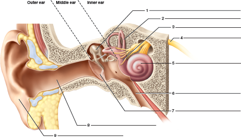
36 identify all indicated structures and ear regions in the following diagram
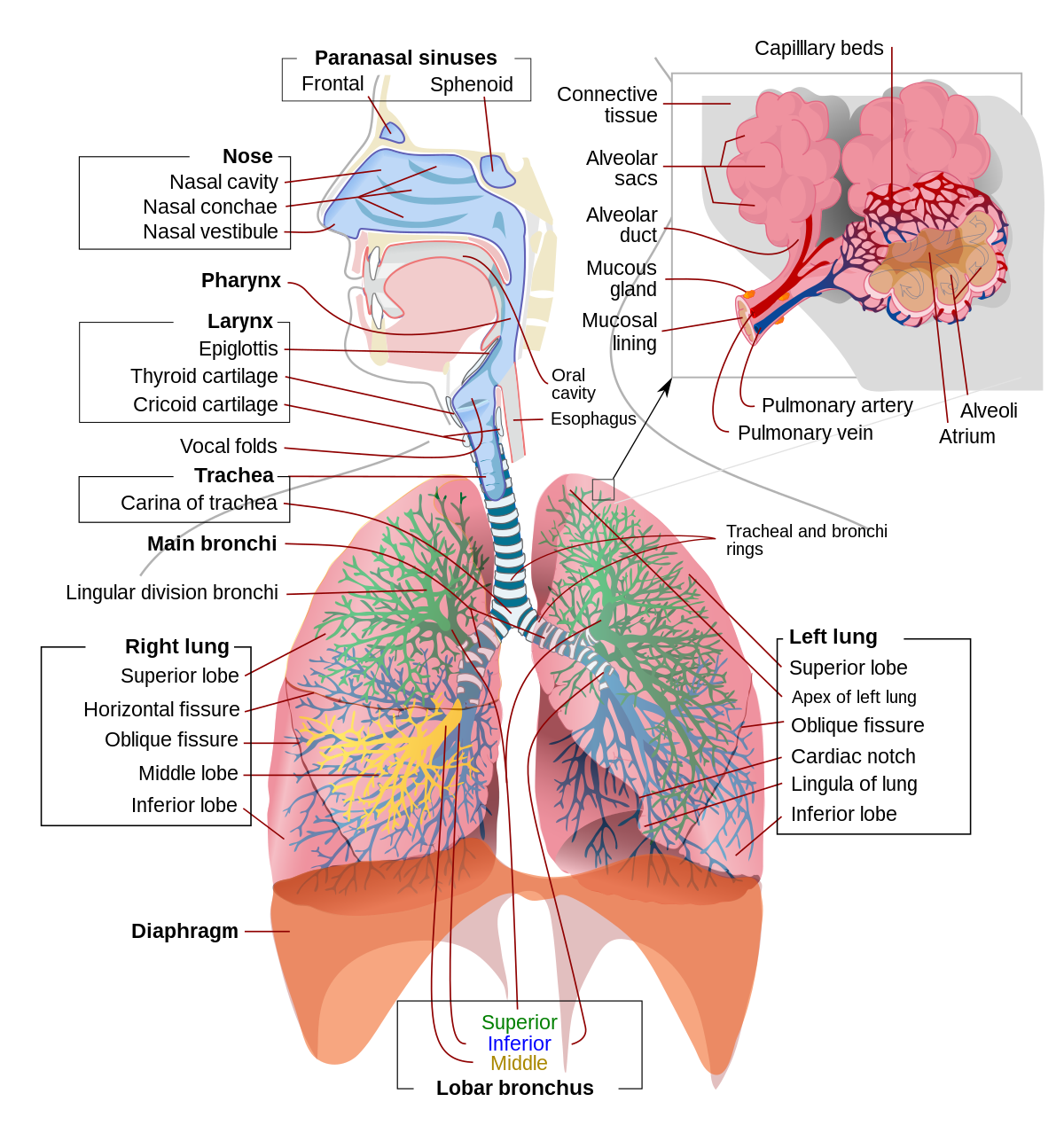
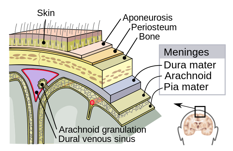
In humans and other mammals, the anatomy of a typical respiratory system is the respiratory tract.The tract is divided into an upper and a lower respiratory tract.The upper tract includes the nose, nasal cavities, sinuses, pharynx and the part of the larynx above the vocal folds.The lower tract (Fig. 2.) includes the lower part of the larynx, the trachea, bronchi, bronchioles and the alveoli. This diagram shows basically what the human brain looks like if it was cut in half just about in the middle between the right and left sides, so that Nrr can see what the structures in the middle ...
Artificial Intelligence Midterm Exam. 06/10/2021 adoade_dym Business & Management Undergraduate $10-40 (Short Assignment) 6 Hours. You must take the online exam on July 28 between 9am and 5pm. It is open book and open note. You will have 2 hours to take the exam once you begin.

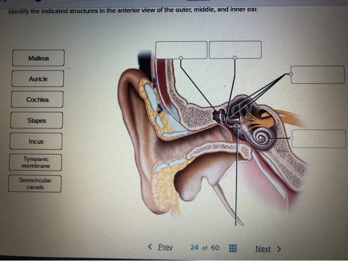
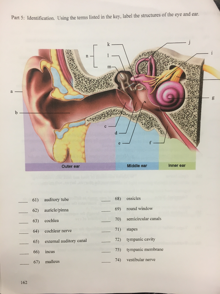
Identify all indicated structures and ear regions in the following diagram
Image: Anatomy of the heart labeled diagram showing the main cardiac structures including the pulmonary artery. Pulmonary Veins The pulmonary veins are responsible for carrying oxygenated blood from the lungs to the left side of the heart, specifically the left atrium (better seen in the diagram at the end of the post). Human Eye Diagram: Contrary to popular belief, the eyes are not perfectly spherical; instead, it is made up of two separate segments fused together. Explore: Facts About The Eye To understand more in detail about our eye and how our eye functions, we need to look into the structure of the human eye. In medical and scientific fields, specific terminology, such as anterior and posterior, conveys the location and directional information of anatomical parts.
Identify all indicated structures and ear regions in the following diagram. Axial Skeleton. The axial skeleton, shown in blue in Figure \(\PageIndex{2}\), consists of a total of 80 bones. Besides the skull, it includes the rib cage and vertebral column. It also includes the three tiny ossicles (hammer, anvil, and stirrup) in the middle ear and the hyoid bone in the throat, to which the tongue and some other soft tissues are attached. Endocrine Glands Diagram As you can see in the image above, men and women share all of the same endocrine glands, besides the reproductive organs. While reproductive organs have the primary function of creating and releasing gametes , they also release a number of hormones which affect the body in different ways. This diagram shows how sound waves travel through the ear, and each step ... As stated above, a given region of the basilar membrane will only move if the ... Several aspects of the phase diagram of water at high pressure are immensely controversial: the location of the melting line 5,10,11,12,13,14,15,16,17,18 and the existence, structure, physical ...
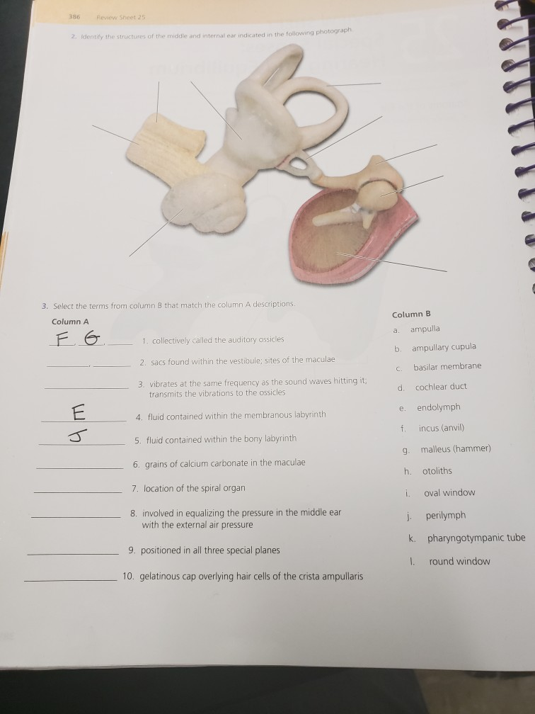
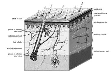
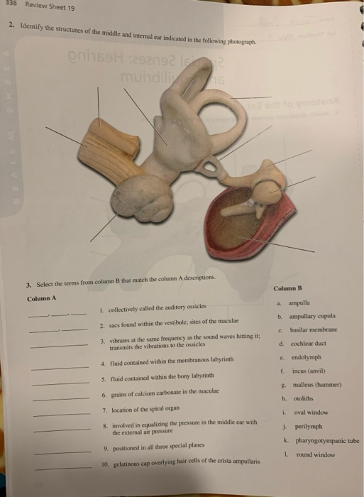
Identify all indicated structures and ear regions in the following photograph. 338 Review Sheet 19 2. Identify the structures of the middle and internal ear ...1 answer · 0 votes: Chegg Anatomy : Time remaining: 01:54:42 Special Senses: Hearing and Equilibrium Anatomy of the Ear 1. Identify all indicated structures and ear regions ... Animal cell size and shape. Animal cells come in all kinds of shapes and sizes, with their size ranging from a few millimeters to micrometers. The largest animal cell is the ostrich egg which has a 5-inch diameter, weighing about 1.2-1.4 kg and the smallest animal cells are the neurons of about 100 microns in diameter. The dermis is the middle layer of the three layers of skin. It's located between the epidermis and the subcutaneous tissue. It contains connective tissue, blood capillaries, oil and sweat glands, nerve endings, and hair follicles. The dermis is split into two parts—the papillary dermis, which is the thin, upper layer, and the reticular dermis ... 21. Identify all indicated structures and ear regions that are provided with leader lines or brackets in the following diagram. PINNA. INNER EAR. EAR.7 pages
The oropharynx is made up of four different regions: Parts of the throat diagram. Parts of a throat pharynx the elongated muscular tube connecting the back of the nose into the neck is known as pharynx. Functions of the pharynx include. To see the illustration of those parts take a look at the following throat diagram. The dermis is a connective tissue layer sandwiched between the epidermis and subcutaneous tissue. The dermis is a fibrous structure composed of collagen, elastic tissue, and other extracellular components that includes vasculature, nerve endings, hair follicles, and glands. The role of the dermis is to support and protect the skin and deeper layers, assist in thermoregulation, and aid in ... Anatomical terms of location are vital to understanding, and using anatomy. They help to avoid any ambiguity that can arise when describing the location of structures. Learning these terms can seem a bit like a foreign language to being with, but they quickly become second nature. Anatomy. The medulla oblongata is one of the three parts of the brainstem, along with the midbrain and the pons. These three collaborating structures are located in front of the cerebellum at the base of the brain and connect to the spinal cord. 1 . Made up of both white and gray matter, the cone-shaped medulla oblongata is formed about 20 ...
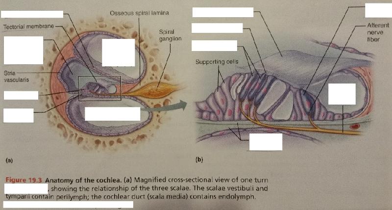
The Organ of Corti is an organ of the inner ear located within the cochlea which contributes to audition. The Organ of Corti includes three rows of outer hair cells and one row of inner hair cells. Vibrations caused by sound waves bend the stereocilia on these hair cells via an electromechanical force. The hair cells convert mechanical energy into electrical energy that is transmitted to the ...

Full labeled anatomical diagrams - Anatomy of the abdomen and digestive system: these general diagrams show the digestive system, with the major human anatomical structures labeled (mouth, tongue, oral cavity, teeth, buccal glands, throat, pharynx, oesophagus, stomach, small intestine, large intestine, liver, gall bladder and pancreas).
Structure and Function of the Inner Ear. The inner ear is entirely enclosed within the temporal bone. It has three separate regions: the cochlea, which is ...

The brainstem is an area located at the base of the brain that contains structures vital for involuntary functions such as the heartbeat and breathing. The brain stem is comprised of the midbrain, pons, and medulla. Midbrain . The midbrain is often considered the smallest region of the brain. It acts as a sort of relay station for auditory and ...
This human anatomy module is about the cranial nerves. It consists of 15 vector anatomical drawings with 280 anatomical structures labeled. It is intended for the use of medical students working on human anatomy, student nurses, physiotherapists, electro-radiological technicians and residents - especially those working in neurology, neurosurgery, otolaryngology - and for any physician ...

The dorsal cavity is at the posterior, or back, of the body, including both the head and the back of the trunk. The dorsal cavity is subdivided into the cranial and spinal cavities. The cranial cavity fills most of the upper part of the skull and contains the brain. The spinal cavity is a very long, narrow cavity inside the vertebral column.
For any palpable lymph node, it's important to assess the following characteristics to help narrow the differential diagnosis: Site: assess the lymph node's location in relation to other anatomical structures. Size: assess the size of the lymph node. Shape: assess the lymph node's borders to determine if they feel regular or irregular.
Trehalose-6-phosphate synthase (TPS) is significant in the growth, development and stress resistance of plants. We identified the cucumber TPS family and its physicochemical properties, domains, gene structures, evolutionary relationships, gene locations, cis-acting elements, conserved motifs, and expression patterns using bioinformatics. Our results uncovered seven CsTPS genes in the cucumber ...
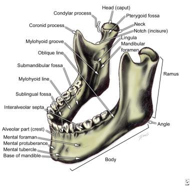
The temporal bone contributes to the lower lateral walls of the skull. It contains the middle and inner portions of the ear, and is crossed by the majority of the cranial nerves. The lower portion of the bone articulates with the mandible, forming the temporomandibular joint of the jaw. In this article, we shall look at the different parts of the temporal bone, their articulations, and any ...

Kidney Structures and Functions Explained (with Picture and Video) Your kidneys are paired organs found on each side of the back portion of the abdominal cavity. The larger left kidney is located a bit higher than the right kidney. Unlike other organs found in the abdomen, the kidneys are located behind the lining (peritoneum) of the abdominal ...
2,683 labeled brain anatomy stock photos, vectors, and illustrations are available royalty-free. See labeled brain anatomy stock video clips. of 27. brain diagram with labels hypothalamus vector brain diagram pons cerebrum and cerebellum brain pons brain anatomy amygdala brain labelled amygdala brain human midbrain diagram pons.

Image: Identify all indicated structures and ear regions ... Indicate whether the following conditions relate to conduction or sensorineural deafess.
Regions of the head and neck. The head is the superior part of the body that is attached to the trunk by the neck. It is the control and communication center as well as the "loading dock" for the body. It houses the brain and therefore is the site of our consciousness: ideas, creativity, imagination, responses, decision making and memory.

Identify all indicated structures and ear regions in the following photograph. 338 Review Sheet 19 2. Identify the structures of the middle and internal ear ...

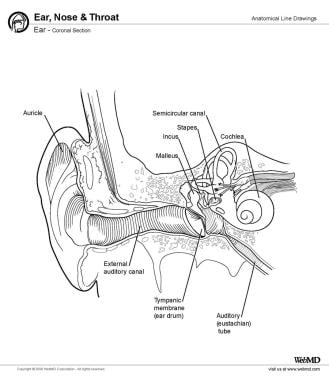
In this article, we'll discuss the auditory ossicles, namely the malleus, incus, and stapes. Inside of the middle ear are the smallest bones in the body-the auditory ossicles, or ear bones. By definition, these three bones are named after their shape: malleus ("hammer"), incus (anvil), and stapes (stirrup). During development, the auditory ossicles are the first bones to fully ossify and ...
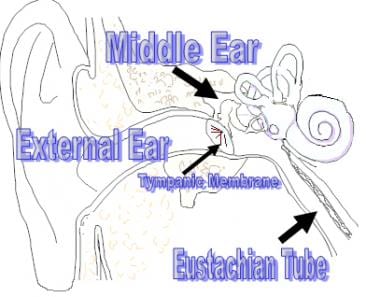
Each section performs a different role in transmitting sound waves to the brain. Outer ear; Middle ear; Inner ear. View the diagrams below to learn more about ...

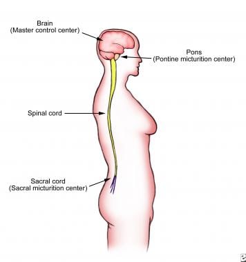
The PNS is all the nerves that branch out from the CNS components and extend to other parts of the body - to the sense organs, muscles, and glands. The PNS connects the CNS to the rest of the body. The primary function of the peripheral nervous system is to connect the brain and spinal cord to the rest of the body and the external environment.
External auditory canal or tube. This is the tube that connects the outer ear to the inside or middle ear. Tympanic membrane (eardrum). The tympanic membrane ...

Four Chambers of the Heart and Blood Circulation. The shape of the human heart is like an upside-down pear, weighing between 7-15 ounces, and is little larger than the size of the fist. It is located between the lungs, in the middle of the chest, behind and slightly to the left of the breast bone. The heart, one of the most significant organs ...
In medical and scientific fields, specific terminology, such as anterior and posterior, conveys the location and directional information of anatomical parts.
Human Eye Diagram: Contrary to popular belief, the eyes are not perfectly spherical; instead, it is made up of two separate segments fused together. Explore: Facts About The Eye To understand more in detail about our eye and how our eye functions, we need to look into the structure of the human eye.

Image: Anatomy of the heart labeled diagram showing the main cardiac structures including the pulmonary artery. Pulmonary Veins The pulmonary veins are responsible for carrying oxygenated blood from the lungs to the left side of the heart, specifically the left atrium (better seen in the diagram at the end of the post).


0 Response to "36 identify all indicated structures and ear regions in the following diagram"
Post a Comment