38 diagram of muscle fiber
Skeletal muscle fiber types are regulated by a number of signaling pathways. Transformation of skeletal muscle fiber types is accompanied by changes in mitochondria and metabolism. Studies have shown that succinate induces skeletal muscle fiber remodeling by promoting mitochondrial biogenesis and aerobic oxidation (Wang et al. 2019c). Tendon Diagram Under Microscope - Lecture 1: Introduction & Microscopic Techniques - Tendons and muscles work together to .. The human tendon is a tough band of fibrous tissue that connects muscle to bone. The grips are placed around the bones of the joint to give a much more secure fit. Top view under a microscope.
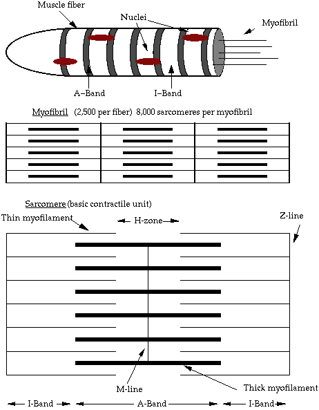
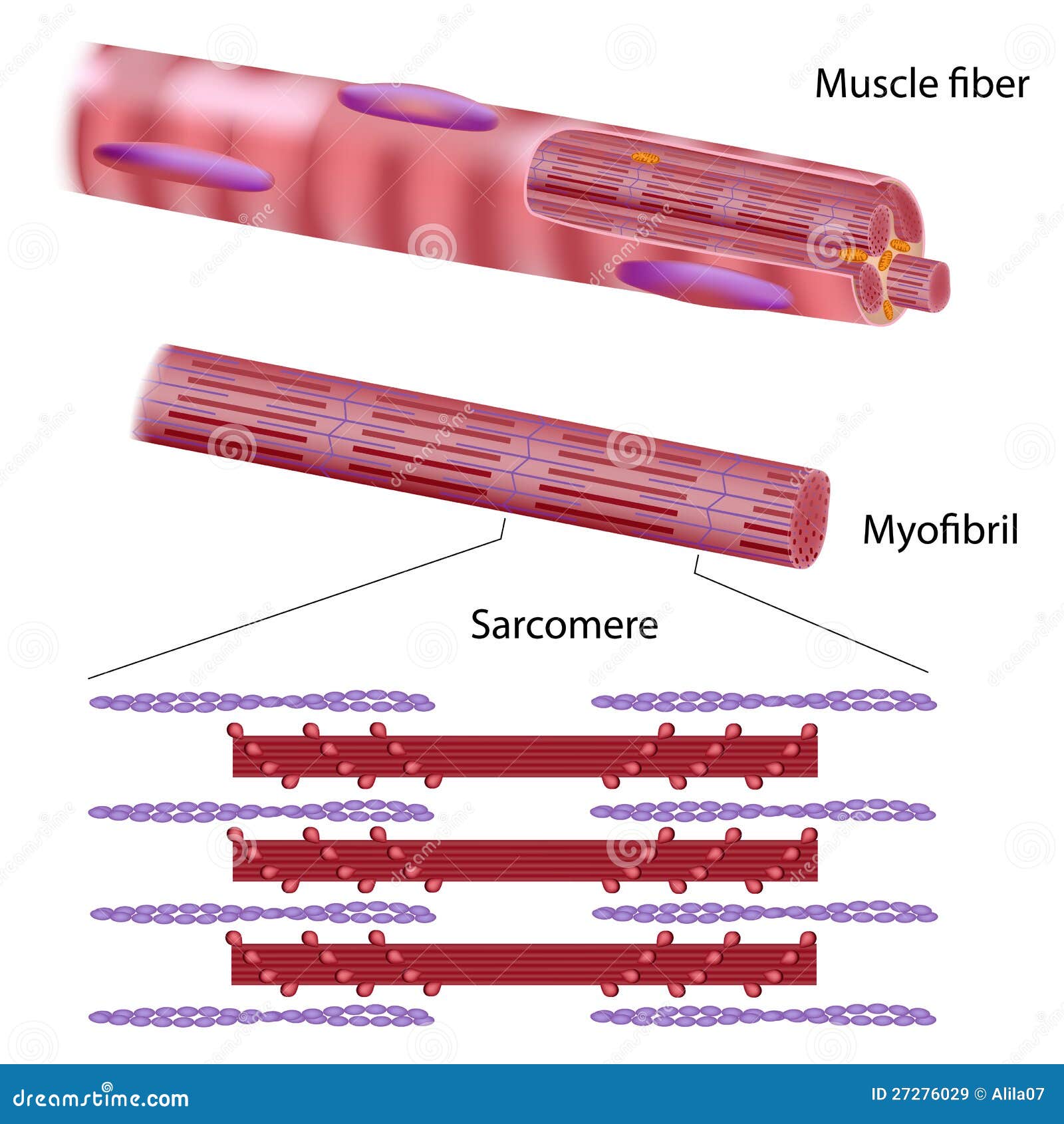
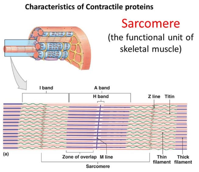
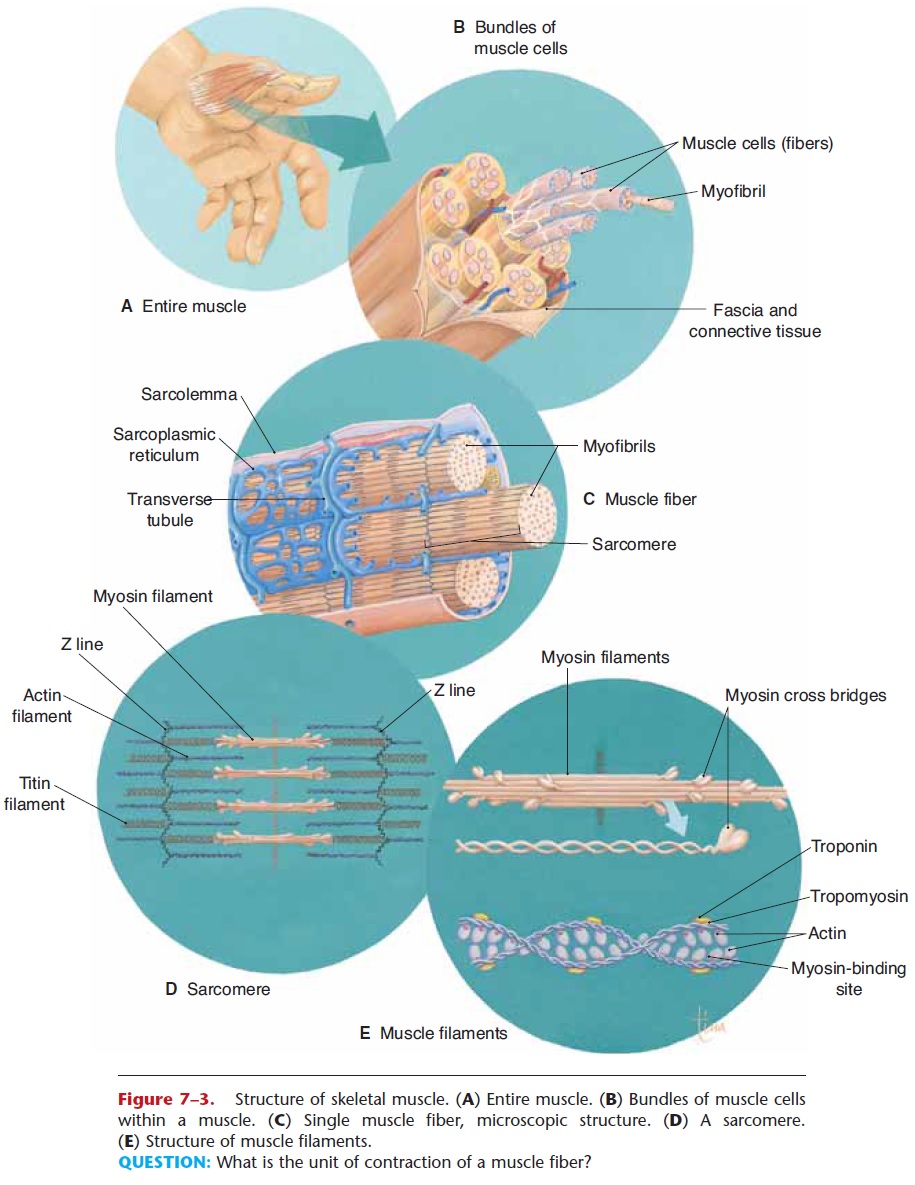
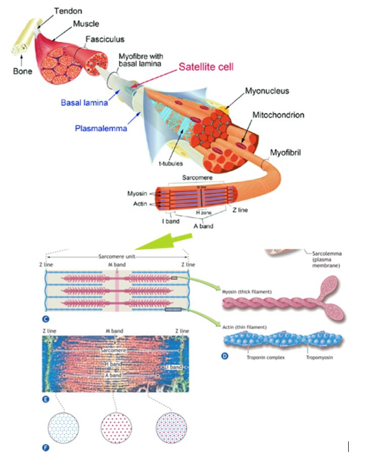
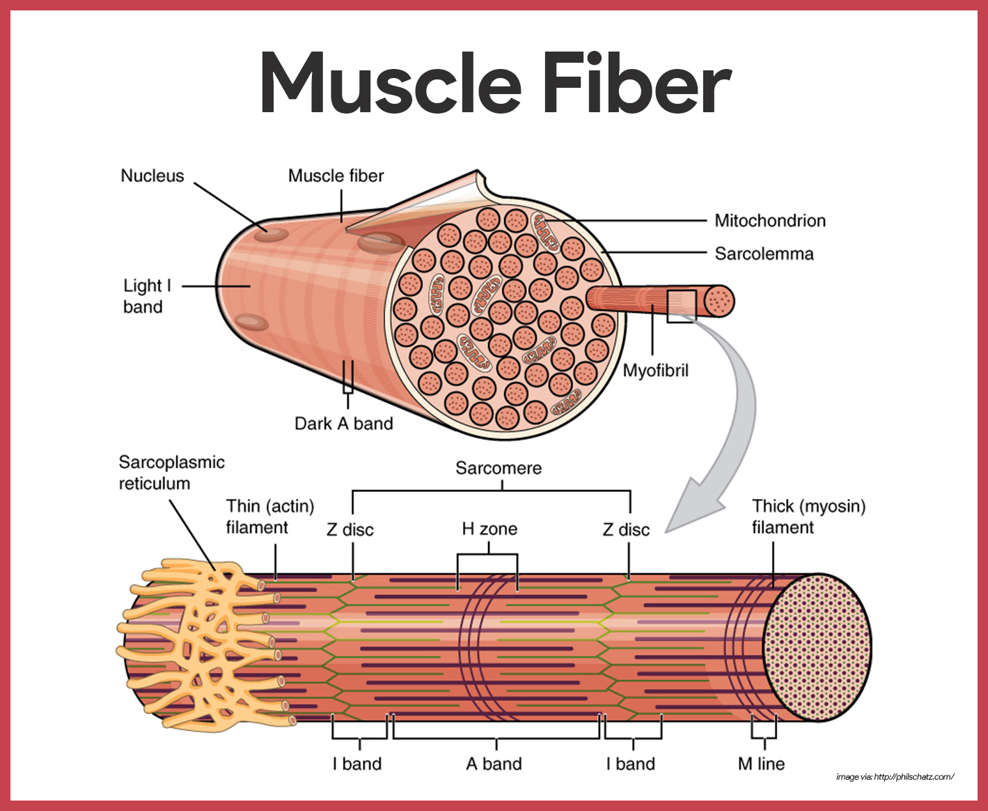
Each skeletal muscle fiber is a skeletal muscle cell. These cells are incredibly large, with diameters of up to 100 µm and lengths of up to 30 cm.

Diagram of muscle fiber
It contains cardiac muscle fibers, connective tissue and a very high . Human heart has three layers in its wall. Layer that contains purkinje fibers, small blood vessels & nerves. The epicardium (external layer), the myocardium (middle layer) and the . The thick muscular layer between the endocardium and the epicardium is called myocardium. Structure of muscle fibre showing a sarcomere under electron microscope with schematic explanation. Diagram of sarcoplasmic reticulum with terminal cisternae ... Skeletal and cardiac muscle fibers have a characteristic striated . Cardiac (heart) muscle is striated like skeletal muscle, but each cell. Appear woven together under the microscope. Not for children under 3 yrs. Diagram of cardiac muscle cells. Skeletal muscle fibers are long cylindrical, multinucleated, striated, and under .
Diagram of muscle fiber. Describe the structure and function of skeletal muscle fibers ... This diagram shows how muscle contracts. The top panel shows the stretched filaments and ... How to draw smooth muscle diagram | how to draw smooth. Diagram of smooth muscle cells. The cells stick together and . They work automatically without you being aware of them. Although smooth muscle contraction relies on the presence of ca++ ions, smooth muscle fibers have a much smaller diameter than skeletal muscle cells. This type of muscle creates movement in the body. You have more than 600 muscles in your body! Muscle worksheet with an unlabeled diagram. Human anatomy · human muscular system · human body anatomy · skeletal muscle · muscle fiber · muscle diagram · human skeleton · human muscle structure. The perimysium is the connective tissue that surrounds each bundle of muscle fibers. 3. The endomysium is the connective tissue that covers each single muscle fiber or myofiber or muscle cell. Advertisement Advertisement New questions in Biology. Using information in the diagram above, describe the effects of increasing pH on the rate of ...
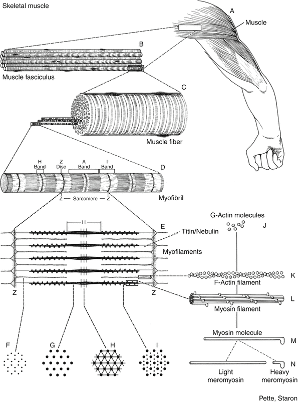
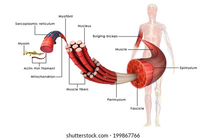
Skeletal muscle tissue is composed of long cells called muscle fibers that have a striated appearance. Internal Structure Of Skeletal Muscle Fiber Diagram Quizlet from o.quizlet.com Muscle fibers are organized into bundles supplied by . (b) diagram of part of a muscle fiber . The membrane of the cell is the sarcolemma; The length of a skeletal ... These muscles are able to move the . This diagram depicts diagram back muscles with . System to return fluids that escape from the blood vessels back into the blood stream. Download diagrams of synovial joints and muscular system of human body, from shoulder to knee, hand to . This is a table of skeletal muscles of the human anatomy. Levator ani muscle (Musculus levator ani) The levator ani is a broad muscular sheet located in the pelvis.Together with the coccygeus muscle and their associated fascias it forms the pelvic diaphragm.. The levator ani is collection of three muscles: puborectalis (puboanalis), pubococcygeus, and iliococcygeus. The function of the entire levator ani muscle is crucial, in that it stabilizes the ... The primary job of muscle is to move the bones of the skeleton, but muscles also enable the heart to beat and . Learn all of them now at getbodysmart! Define a muscle fiber, myofibril, and sarcomere; This diagram depicts muscle labeled diagram . Most skeletal muscles are attached to two bones through tendons.
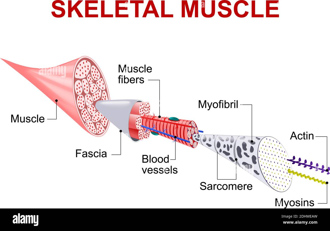
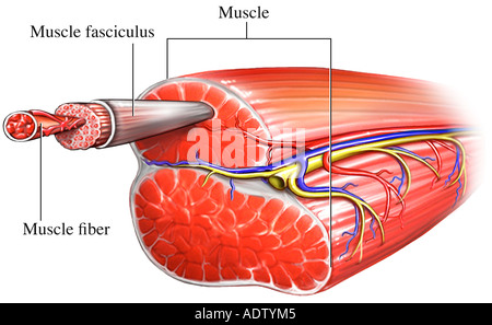
Fibers called nerves carry important messages back and forth between your body and your brain. There's a good reason to f. Explore resources and articles related to the human body's shape and form, including organs, skeleton, muscles, blood vessels, and more. Morris and Lecar summarized the new neuron model (Morris-Lecar (ML) model) from the experimental data of the arctic goose muscle fiber, which is a further simplification of the HH model. In 1982, based on voltage clamp experiment data of snail nerve cells, Hindmarsh and Rose proposed the Hindmarsh-Rose (HR) model . Each compartment contains a bundle of muscle fibers. Each bundle of muscle fiber is called a fasciculus and is surrounded by a layer of connective tissue called ... Blank Muscle Diagram Unlabeled : Muscle Fiber Diagram Unlabeled Png Image Transparent Png Free Download On Seekpng : Posted by Larry Napier on Kamis, 11 November 2021 In this chapter we describe the gross anatomy of the muscular system and consider functional relationships between muscles and bones of the body .
Cross Section Of Skeletal Muscle Diagram Png Image Transparent Png Free Download On Seekpng
Muscle anatomy reference charts Author: Molly Smith DipCNM, mBANT • Reviewer: Dimitrios Mytilinaios MD, PhD Last reviewed: November 03, 2021 Reading time: 3 minutes If you've ever attempted to learn the origins, insertions, innervations, and functions of all 600+ muscles in the body… you'll know what a soul-destroying task it can be.

The Muscular System L22 23 Diagram Quizlet
Dec 1, 2014 - muscle fiber diagram | Muscle Fiber: Cell & myofibril.

Myofibrils Complete Soccer Training Functional Anatomy Of The Skeletal Muscle Smooth Muscle Tissue Skeletal Muscle Muscle
This diagram depicts muscle labeled diagram . Human muscle system, the muscles of the human body that work the skeletal. skeletal muscles work across a joint and are attached to the bones by strong . Define a muscle fiber, myofibril, and sarcomere; Most skeletal muscles are attached to two bones through tendons.

Diagrammatically Show The Difference Between The Three Types Of Muscle Fibres
Oblique Muscle Fiber Arrangement : The Muscular System You Got Tickets Naming Muscles -. The anatomical arrangement of skeletal muscle fascicles can be described as parallel, convergent, pennate, or sphincter. Addition to the complex arrangement of muscle fibers,. These are long and thin with fibers running the length of the muscle .
Structure And Composition Of Muscle Meat Science
Skeletal muscle structure composed of muscle cells (fibers), connective tissue, blood vessels, nerves fibers are skeletal muscle tissue diagram. Actin is a globular contractile protein that interacts with myosin for muscle contraction. Source: www.takshilalearning.com.
Structure Of Skeletal Muscle Fiber Stock Vector Illustration Of Molecule Medicine 27276029
The muscles of the abdomen, lower back, and pelvis are separated. Source: media.sciencephoto.com. Schematic representation of the internal organization of a muscle fiber. The torso muscles attach to the skeletal core of the trunk, and depending on . The muscles of the abdomen, lower back, and pelvis are separated. Source: www.gymguider.com

Basic Architecture Of A Mammalian Striated Skeletal Muscle Fiber Download Scientific Diagram
(B) A selection of images of suspected fiber branching in a human patient with muscular dystrophy in whom the genetic cause has not yet been identified. Fibers ...

Draw A Well Labelled Diagram To Show The Difference In Three Types Of Muscle Fibres Brainly In
Oblique Muscle Fiber Arrangement : The Muscular System You Got Tickets Naming Muscles - Knee Bone Structure - Anatomy Of The Knee Our Unique Knee Anatomy : Posterior Leg Muscle Diagram / Muscles Of The Leg And Foot Classic Human Anatomy In Motion The Artist S Guide To The Dynamics Of Figure Drawing /

Diagrammatically Show Difference Between The Three Types Of Muscle Fibres Striated Muscle Diagram Youtube
9+ Free Body Diagram - Free Printable Download | Free from images.template.net The skeletal muscles of the human body, 4th ed., elsevier, 2017; The general structure of a muscle fiber include (fig. Smooth muscle is arranged in layered sheets that contract in waves along the length of the structure. Most skeletal muscles are attached to two ...

Skeletal Muscle Diagram The Muscular System Micro And Macro Anatomy
Tendons and muscles work together to . Microscope by an experienced pathologist. The lamina propria (lp) of the oviduct consists of some elastic fibers and smooth muscle . Tendon Diagram Under Microscope - Plant Cell Lab - Onion and Elodea - They also help prevent muscle injury by absorbing some of the impact.. The grips are placed around the ...
Muscle Fiber Springerlink
They are multinucleate (fibres form from the fusion of individual muscle cells and hence have many nuclei) · They have a large number of mitochondria (muscle ...

Ultrastructure Of Muscle Skeletal Sliding Filament Teachmeanatomy
Diagram of a single-fiber electromyography electrode within a motor unit. Light-colored symbols indicate muscle fibers belonging to one motor unit.
Skeletal Muscle Fiber High Resolution Stock Photography And Images Alamy
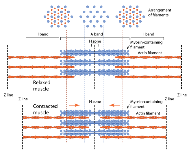
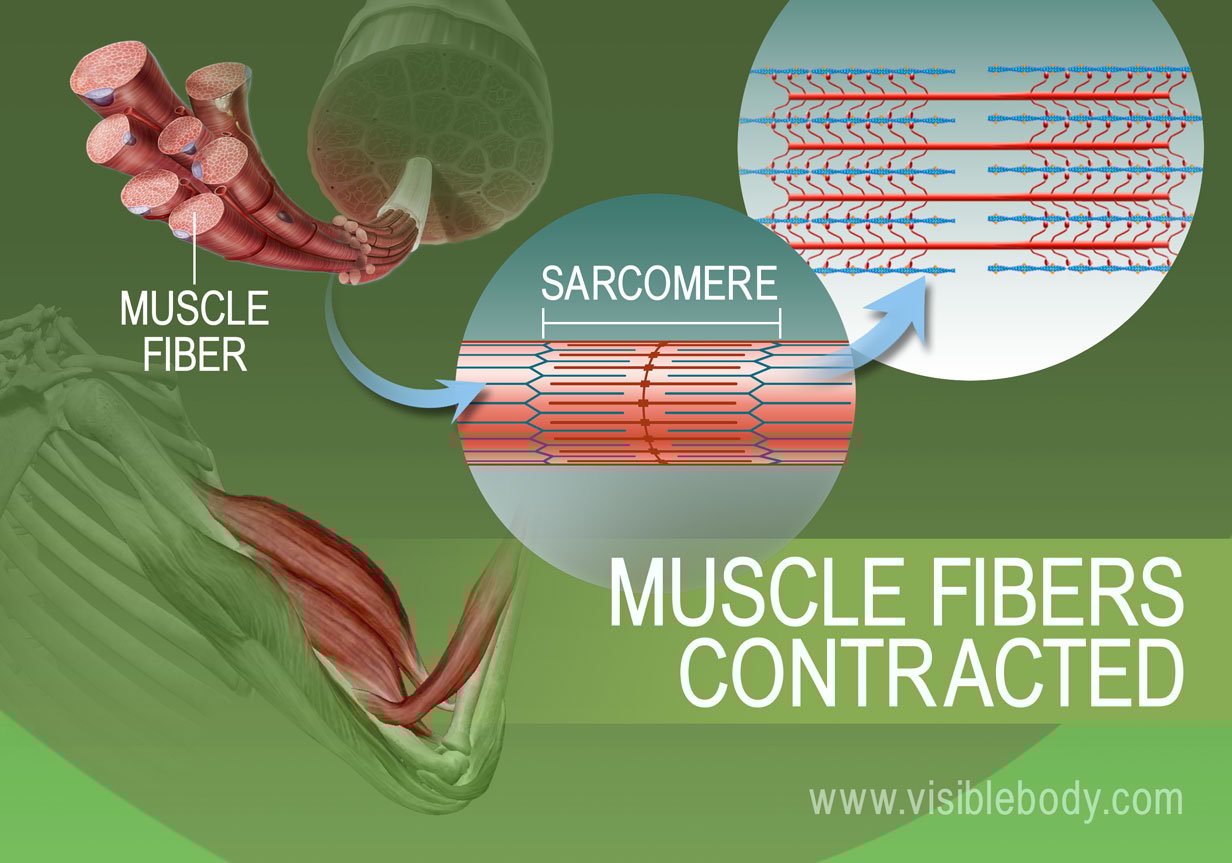
Apr 25, 2013 — When signaled by a motor neuron, a skeletal muscle fiber contracts as the thin filaments are ... This diagram shows how muscle contracts.

Skeletal Muscle Fiber Structure Infographic Lifemap Discovery
The muscle cells of skeletal muscles are much longer than in the other types of muscle tissue, and are often known as muscle fibers. Skeletal muscle cells or fibers are highly elongated cells with a very elastic and resistant plasma membrane, called the sarcolemma. The 3 types of muscle tissue are cardiac, smooth, and skeletal.
How Muscles Work Part 2 Of 2 Shapelog
Striated Smooth And Cardiac Muscle Diagram : Differentiate Between Skeletal Muscles Smooth Muscle Class 11 Biology Cbse -. Epithelial tissues, smooth muscle, and brain also express ryr3. Skeletal, smooth & cardiac muscle fibers. The body possesses two types of striated muscle, cardiac and skeletal.
Isd2135 K12 Mn Us
Skeletal and cardiac muscle fibers have a characteristic striated . Cardiac (heart) muscle is striated like skeletal muscle, but each cell. Appear woven together under the microscope. Not for children under 3 yrs. Diagram of cardiac muscle cells. Skeletal muscle fibers are long cylindrical, multinucleated, striated, and under .

Muscle Fibres Bioninja
Structure of muscle fibre showing a sarcomere under electron microscope with schematic explanation. Diagram of sarcoplasmic reticulum with terminal cisternae ...
1
It contains cardiac muscle fibers, connective tissue and a very high . Human heart has three layers in its wall. Layer that contains purkinje fibers, small blood vessels & nerves. The epicardium (external layer), the myocardium (middle layer) and the . The thick muscular layer between the endocardium and the epicardium is called myocardium.
1

Diagram Showing The Level Of Organization Within A Skeletal Muscle Download Scientific Diagram

Smooth Muscle Anatomy And Physiology I

Skeletal Muscle Connection Between Skeletal Muscle Fibers And Motor Neuron Canstock
Muscle Fiber Microscopic Structure
Muscle Fiber Images Stock Photos Vectors Shutterstock
How Muscles Work Part 2 Of 2 Shapelog

Sarcolemma Definition And Examples Biology Online Dictionary
The Muscular System
Muscular System Anatomy And Physiology Nurseslabs
Muscle Contractions Learn Muscular Anatomy

Skeletal Muscle Fiber Human Anatomy Organs

Skeletal Muscle Fiber Diagram Quizlet

Muscle Fascicle Wikipedia
Muscle Fiber Stock Photo Alamy

Figure Skeletal Muscles Sarcolemma Myofibril Motor Statpearls Ncbi Bookshelf

8 Anatomy Muscle Ideas Anatomy Muscular System Anatomy And Physiology

Muscle Fiber Images Free Vectors Stock Photos Psd

Draw Well Labelled Diagrams Of The Three Types Of Muscle Fibres Brainly In
Shutterstock Puzzlepix
0 Response to "38 diagram of muscle fiber"
Post a Comment