39 gram positive cell wall diagram
Gram-positive cell wall. Gram-positive cell wall is thick measuring about 15-80 nm and more homogenous compared to gram-negative cell wall. This cell wall consists of large amount of peptidoglycan arranged in several layers. Peptidoglycan in gram-positive cell wall constitutes about 40-80% of the dry weight. In electron micrographs, the Gram-positive cell wall appears as a broad, dense wall 20-80 nm thick and consisting of numerous interconnecting layers of peptidoglycan (see Figs. 1A and 1B). Chemically, 60 to 90% of the Gram-positive cell wall is peptidoglycan. In Gram-positive bacteria it is thought that the peptidoglycan is laid down in cables ...
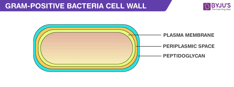
Gram-Positive Cell Wall. The composition of gram-positive bacteria cell wall includes: Peptidoglycan. It is permeable, cross-linked organic polymer and rigid structure which plays an important role in providing shape and strength to the cell wall. It makes up about 90% of the cell wall enclosing the plasma membrane and protects the cell from ...
Gram positive cell wall diagram
The gram-Positive Cell wall of Bacteria Bacterial cell wall that is gram-positive contains peptidoglycan and teichoic acids with some species having additional carbohydrates and proteins. The murein component is what gives shape to the gram-positive bacterial cell wall; it also helps the bacteria cells to resist osmotic pressure. Cell wall structures of Gram-positive and Gram-negative bacteria and fungi. (A) Gram-positive bacteria have a single lipid bilayer surrounded ... The analysis of the mycolic acid as the major characteristic of MbT cell-wall component has been used [19] to distinguish between MbT and strains of Gram-positive (S. aureus) and Gram-negative (K ...
Gram positive cell wall diagram. 4 Bacteria: Cell Walls . It is important to note that not all bacteria have a cell wall.Having said that though, it is also important to note that most bacteria (about 90%) have a cell wall and they typically have one of two types: a gram positive cell wall or a gram negative cell wall.. The two different cell wall types can be identified in the lab by a differential stain known as the Gram stain. Each of the following statements concerning the gram-positive cell wall is true EXCEPT. ... In Figure 4.3, which diagram of a cell wall is a gram-negative cell wall? B. In Figure 4.3, which diagram of a cell wall possesses lipid A/endotoxin responsible for symptoms associated with infection. B. Gram-positive cell walls, once thought to be relatively simple structural entities, can be quite different from one another, especially when cell wall turnover is taken into account (8, 9, 25, 29). The cell walls of gram-negative bacteria follow a more general structural format than that of gram-positive bacteria, which is strictly adhered to ... Important Chemical Components of Surface Structures. Cell Wall Peptidoglycans: Both Gram-positive and Gram-negative bacteria possess cell wall peptidoglycans, which confer the characteristic cell shape and provide the cell with mechanical protection. Peptidoglycans are unique to prokaryotic organisms and consist of a glycan backbone of muramic acid and glucosamine (both N-acetylated), and ...
2) Each of the following statements concerning the gram-positive cell wall is true EXCEPT. A) it maintains the shape of the cell. B) it is sensitive to lysozyme. C) it protects the cell in a hypertonic environment. D) it contains teichoic acids. E) it is sensitive to penicillin. Gram staining Procedure. Retains the crystal violet dye and appear purple in colour. Does not retain the crystal violet dye and appear pink in colour. These differences between the Cell Wall of Gram-positive and Gram-negative Bacteria are classified based on their structure, composition of the cell and by the procedure of Gram staining technique. The Gram-Positive Cell Wall. As mentioned in the previous section on peptidoglycan, Gram-positive bacteria are those that retain the initial dye crystal violet during the Gram stain procedure and appear purple when observed through the microscope. As we will learn in lab, this is a result of the structure and function of the Gram-positive cell wall. Gram positive bacteria. Gram positive bacteria have a distinctive purple appearance when observed under a light microscope following Gram staining. This is due to retention of the purple crystal violet stain in the thick peptidoglycan layer of the cell wall. Examples of Gram positive bacteria include all staphylococci, all streptococci and some ...
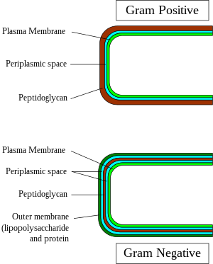
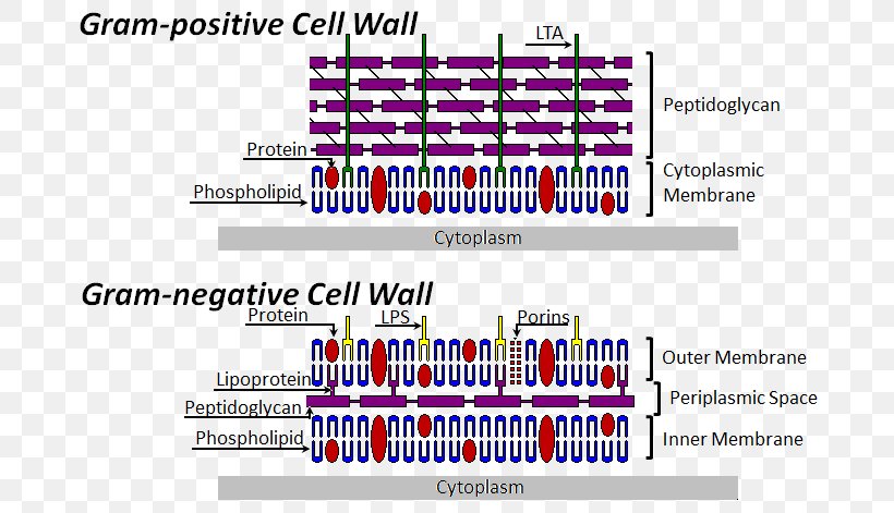
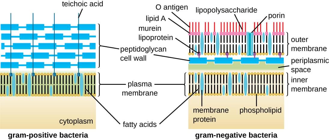
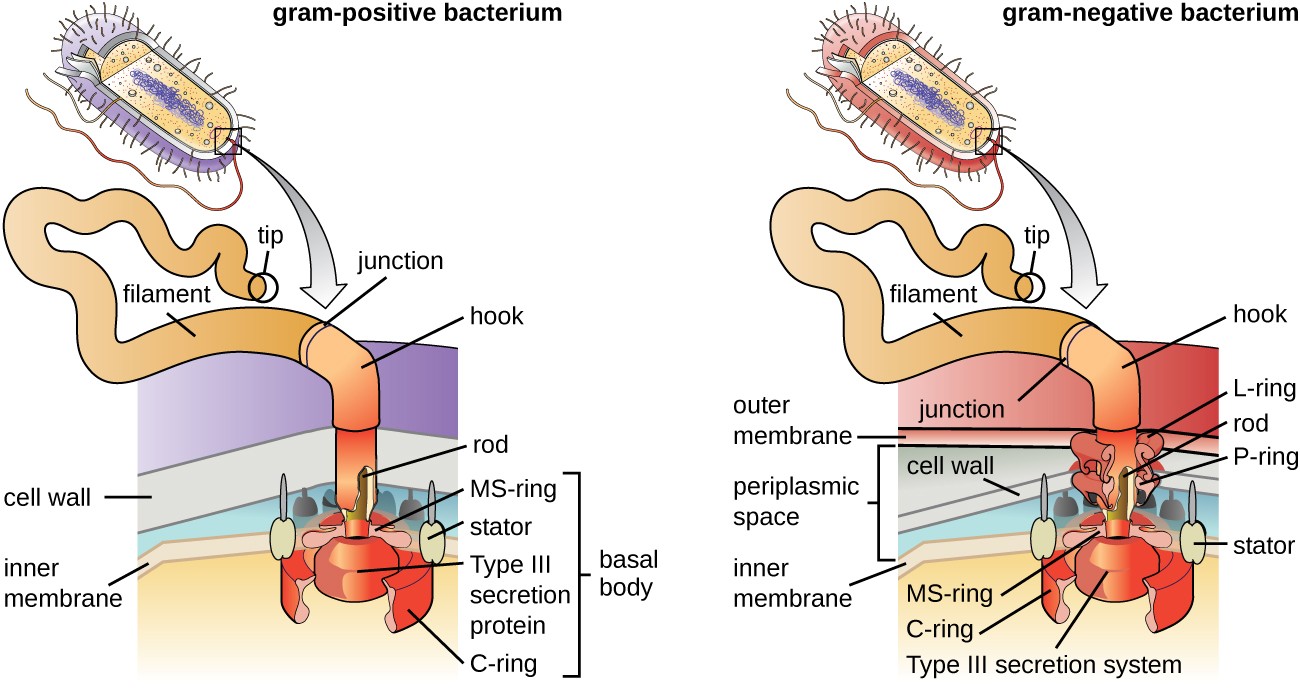
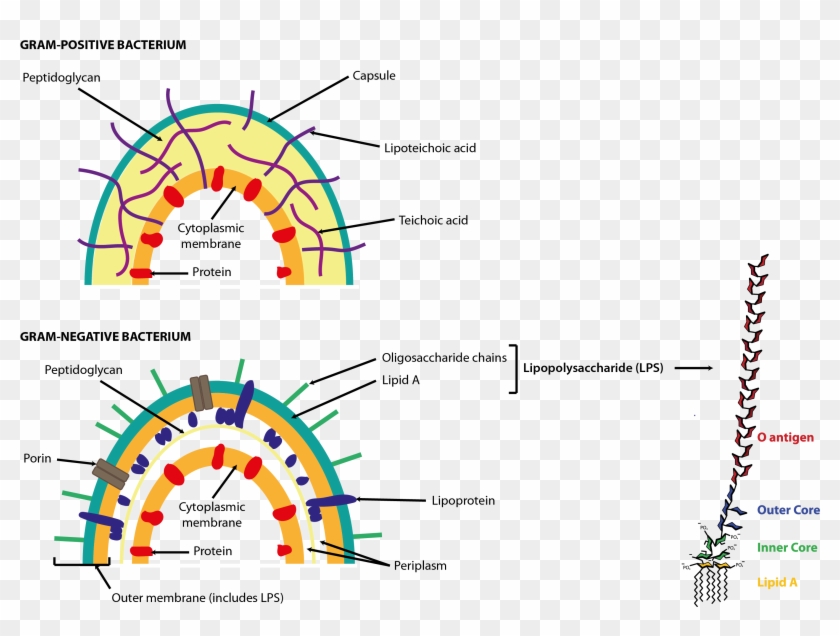
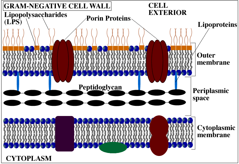
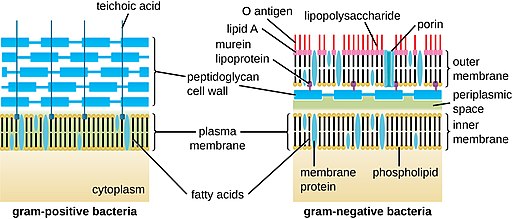
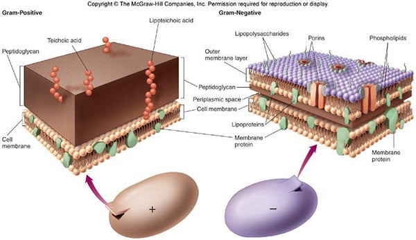
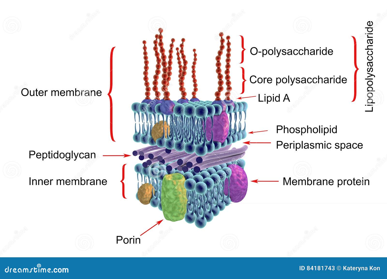
From the peptidoglycan inwards all bacterial cells are very similar. Going further out, the bacterial world divides into two major classes: Gram positive (Gram ... You have isolated a motile, gram-positive cell with no visible nucleus. You can safely assume that the cell. has a cell wall. Fimbriae and pili differ in that pili … In Figure 4.3, which diagram of a cell wall is a gram-negative cell wall (smaller) gram-negative. In Figure 4.3, which diagram of a cell wall is a toxic cell wall (smaller) gram ... Most of the gram positive cell wall contain considerable amount of teichoic acid and teichuronic acid. In addition, they may contain polysaccharide molecules. The gram negative cell wall contains three components that lie outside the peptidoglycan layer: 1. Lipoprotein. 2. Outer membrane and . 3. Lipopolysaccharide. Cell membrane · by M Rajagopal · 2017 · Cited by 123 — The cell envelopes of most bacteria fall into one of two major groups. Gram-negative bacteria have an inner, cytoplasmic membrane surrounded ...MRSA: Methcillin-resistant Staphylococcus aur...PGT: Peptidoglycan glycosyltransferasePG: PeptidoglycanCPS: Capsular polysaccharides
1
Grams positive bacteria are a category of bacteria. Their cell wall is known as gram positive cell wall. This is because it has a thick peptidoglycan layer. It is multilayered and possesses teichoic acids. In grams staining, gram positive cell wall stains in purple colour due to the retention of crystal violet stain.
What Is The Structure And Composition Of A Bacterial Cell Wall Quora
Compare and contrast the cell walls of typical Gram-positive and Gram-negative bacteria. 3. Relate bacterial cell wall structure to the Gram-staining reaction. 37 . 38 Bacterial Cell Wall • Peptidoglycan (murein) -rigid structure that lies just outside the cell plasma membrane
Gram Positive Bacteria Wikipedia
Gram-positive Cell Wall. Gram-negative Cell Wall. Outer Membrane. Cytoplasmic Membrane. Membrane Proteins. Porin. RETURN to CELL ENVELOPE . RETURN to CELL DIAGRAM ...

Peptidoglycans
This quick video describes in detail the cell wall structure of gram positive bacteria.Check out http://eacharya.tumblr.com for more!
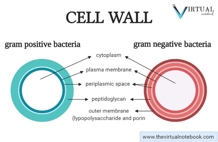
Cell Wall Structure And Composition The Virtual Notebook
The gram-positive cell wall — Types of bacterial cell envelopes[edit]. The gram-positive cell wall[edit]. Schematic of ...
Cell Wall And Cell Membrane
Gram positive bacteria are a group of organisms that fall under the phylum Firmicutes (however, a few species have a Gram negative cell wall structure). As compared to Gram negative bacteria, this group of bacteria is characterized by their ability to retain the primary stain (Crystal violet) during Gram staining (giving a positive result).
2
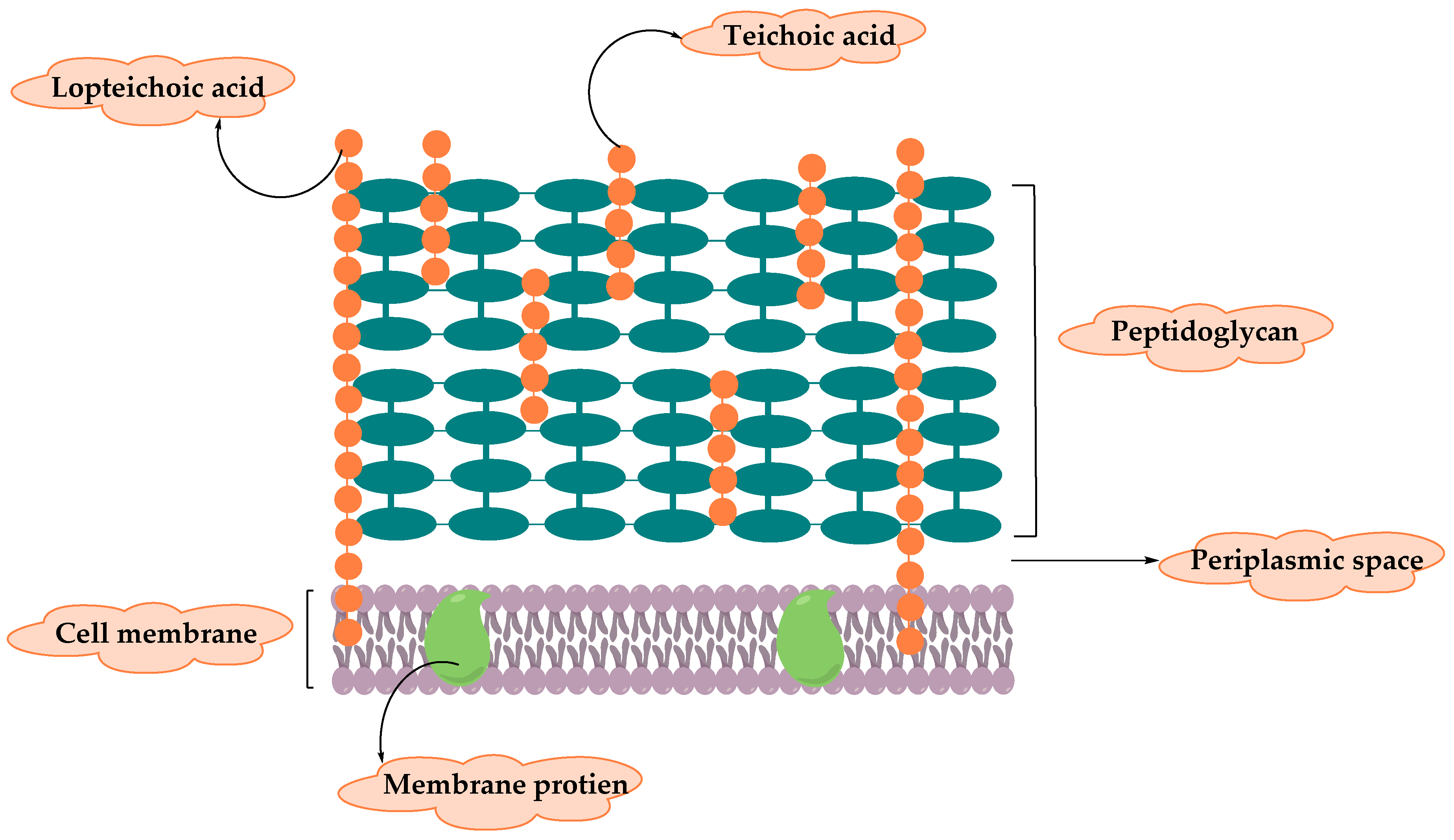
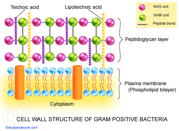
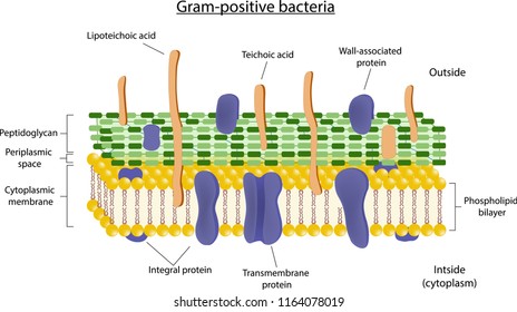
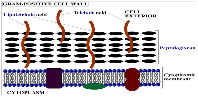
In the Gram-positive Bacteria (those that retain the purple crystal violet dye when subjected to the Gram-staining procedure), the cell wall consists of several layers of peptidoglycan. Running perpendicular to the peptidoglycan sheets is a group of molecules called teichoic acids which are unique to the Gram-positive cell wall (Figure 14).

Schematic Structure Of Gram Positive And Gram Negative Cell Walls Download Scientific Diagram
The cell wall of bacteria is located at the inner side of the capsule. Gram positive and gram negative. The cytoplasm is enclosed by three layers the outermost slime or capsule the middle cell wall and inner cell membrane. It gives shape to the cell. It give shaperigidity and support to the cell.
1
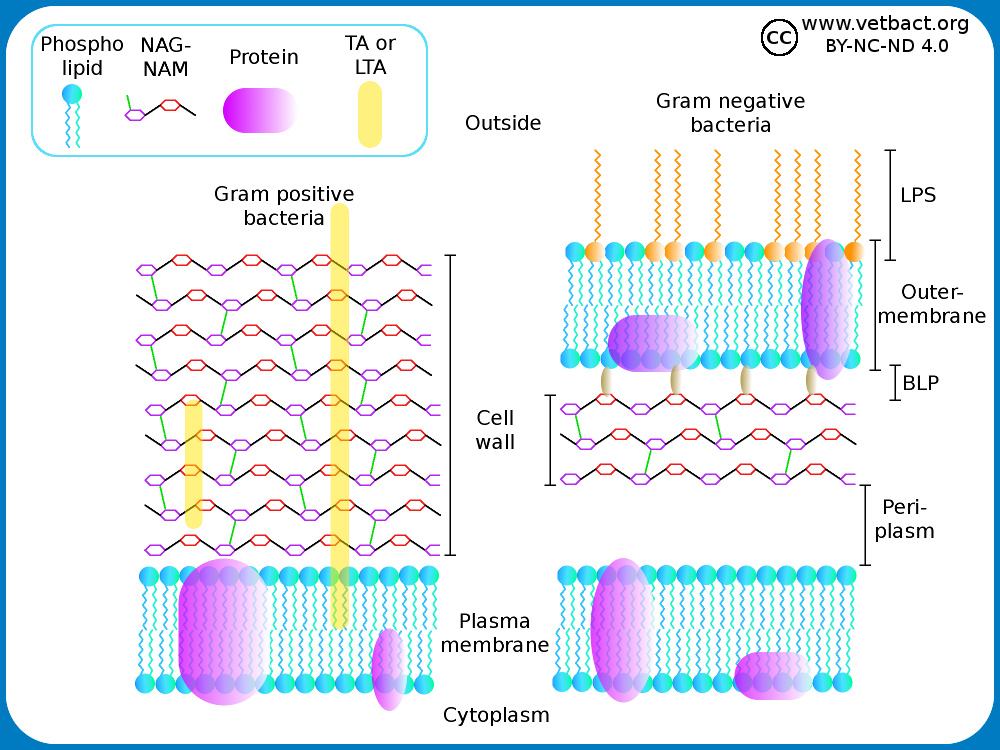
In Gram-positive bacteria, peptidoglycan makes up as much as 90% of the thick cell wall enclosing the plasma membrane. Gram Staining! During Gram staining, these thick, multiple layers (20-80 nm) of peptidoglycan retain the dark purple primary stain crystal violet, whereas Gram-negative bacteria stain pink.

Lysing Enzymes
For the gram-positive cell wall, it has a thickness of about 20-80nm thickness made up of a thick peptidoglycan layer outside its cell membrane, unlike the thin layer of gram-negative bacteria (10-15nm) which has a very thin layer of the peptidoglycan of 2-7nm but has a thicker lipid layer making it quite complex than the Gram-positive cell wall.
Gram Positive Cell Wall Labster Theory
Gram-positive cell wall. The Gram-positive cell wall is thick (15-80 nm) and more homogenous than that of the thin (2 nm) Gram-negative cell wall. The Gram-positive cell wall contains large amount of peptidoglycan present in several layers that constitutes about 40-80% of dry weight of the cell wall.
Bacterial Cell Structure Cell Wall Gram Positive Bacteria Gram Negative Bacteria Png 696x471px Bacterial Cell Structure
Briefly describe how antibiotics such as penicillins, cephalosporins, and vancomycin affect bacteria and relate this to their cell wall synthesis. State what color Gram-positive bacteria stain after Gram staining. State what color Gram-negative bacteria stain after Gram staining. State what color acid-fast bacteria stain after acid-fast staining.

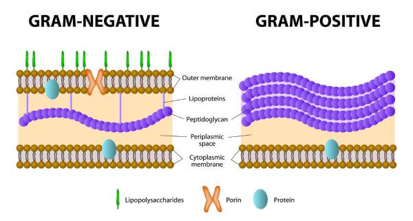
Gram Positive Vs Gram Negative Technology Networks
The results from images shown in e-i support a common process of synthesis and maturation of the cell wall on the spherical parts of Gram-positive bacteria in both spherical and rod-shaped species.
Gram Positive Vs Gram Negative Biology Dictionary
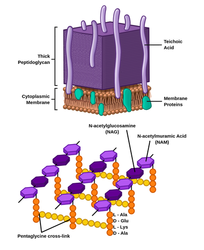
Techoic acids consist of. 2 classes of techoic acids. Many layers of peptidoglycan, forming a thick, rigid structure. In addition to peptidoglycan, techoic acids and phosphate. Consists primarily of an alcohol. lipotechoic acid & peptidoglycan. Cell wall of gram-positive bacteria.
Molecules Free Full Text Resistance Of Gram Positive Bacteria To Current Antibacterial Agents And Overcoming Approaches Html
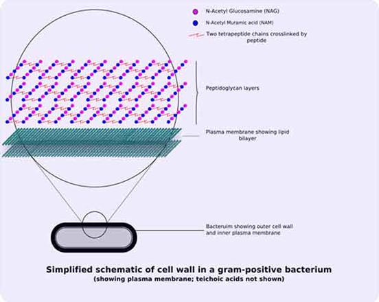
Gram Stain Mechanism: Gram Positive Cell Wall: Gram-positive bacteria have a thick mesh-like cell wall which is made up of peptidoglycan (50-90% of cell wall), which stains purple. Peptidoglycan is mainly a polysaccharide composed of two subunits called N-acetyl glucosamine and N-acetyl muramic acid.
Bacterial Cell Wall Targets Identification Creative Biolabs
The analysis of the mycolic acid as the major characteristic of MbT cell-wall component has been used [19] to distinguish between MbT and strains of Gram-positive (S. aureus) and Gram-negative (K ...

Pathogen Recognition And Innate Immunity Cell
Cell wall structures of Gram-positive and Gram-negative bacteria and fungi. (A) Gram-positive bacteria have a single lipid bilayer surrounded ...
Unique Characteristics Of Prokaryotic Cells Microbiology
The gram-Positive Cell wall of Bacteria Bacterial cell wall that is gram-positive contains peptidoglycan and teichoic acids with some species having additional carbohydrates and proteins. The murein component is what gives shape to the gram-positive bacterial cell wall; it also helps the bacteria cells to resist osmotic pressure.
Cell Walls Of Gram Positive And Gram Negative Bacteria Lipopolysaccharide In Bacteria Clipart 216045 Pikpng
Cell Wall Of Bacteria Structure Functions Gram Positive And Gram Negative Cell Walls Study Score

Microbiological Educational Diagram Sample Cell Envelope Of Gram Positive Bacteria Microbiology Education Positivity
Gram Positive Bacteria Characteristics And Structure
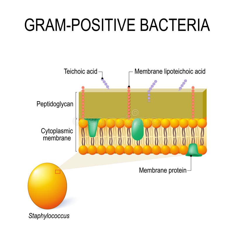
Cell Wall Structure Of Gram Positive Bacteria For Example Staphylococcus Stock Vector Illustration Of Crystal Bacterial 125533441

Gram Positive Vs Gram Negative Cell Walls Cell Wall Plasma Membrane Biology Notes

Gram Positive Vs Gram Negative Bacteria Microbe Online
Gram Positive Cell Images Stock Photos Vectors Shutterstock
The Virtual Edge

Gram Positive Cell Wall Ppt Download

Bacterial Pathogens Research

Bacteria Cell Wall Gram Positive And Gram Negative
Cell Wall Gram Positive Vs Negative Bacteria Easy Biology Class
Differences Between Gram Positive And Gram Negative Bacteria
Bacterial Cell Wall Structure Gram Positive Negative
:max_bytes(150000):strip_icc()/gram_positive_bacteria-5b7f3032c9e77c0050f88457.jpg)
Gram Positive Vs Gram Negative Bacteria

20 Key Difference Between Gram Positive And Gram Negative Bacteria Cell Walls Core Differences
Experiment 4a Lab04 Virtual Edge Molb 2021 College Of Agriculture And Natural Sciences
Gram Negative Bacteria Cell Wall Stock Illustrations 11 Gram Negative Bacteria Cell Wall Stock Illustrations Vectors Clipart Dreamstime

Bacteria 101 Cell Walls Gram Staining Common Pathogens Tusom Pharmwiki

Week 2 How Do Antibiotics Work 4 1 Gram Positive And Gram Negative Bacteria Openlearn Open University Uar 1
0 Response to "39 gram positive cell wall diagram"
Post a Comment