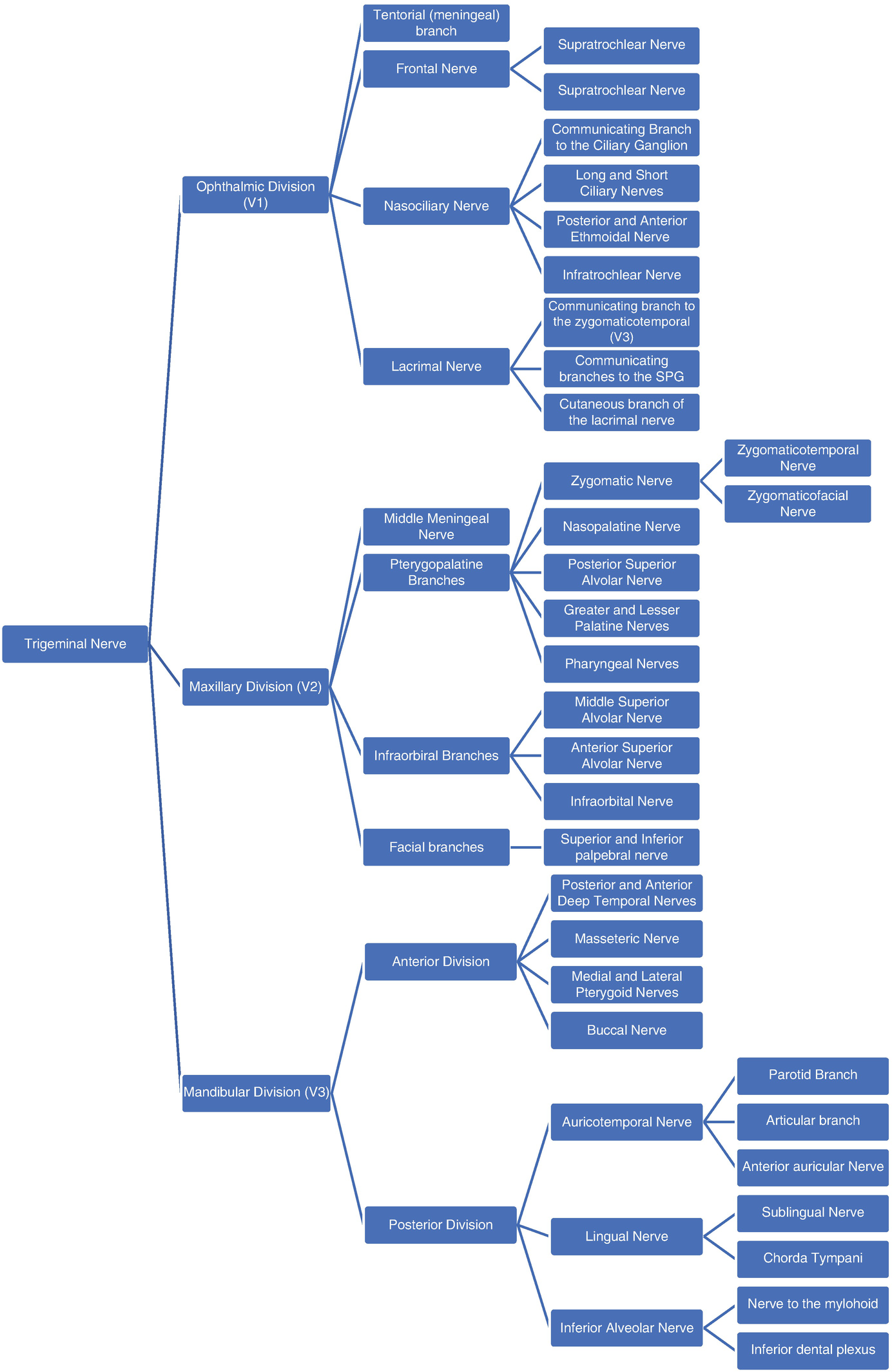
36 trigeminal nerve branches diagram
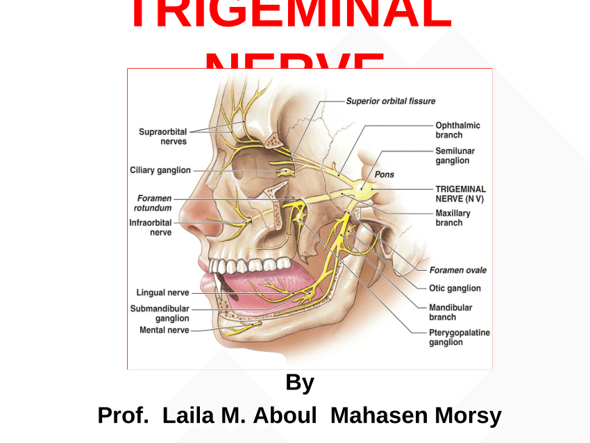
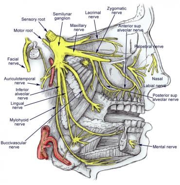
Diagram of the third branch (mandibular) of the trigeminal nerve with its branches. View Media Gallery The mandibular nerve carries sensory information from the lower lip, the lower teeth, gums, the chin and jaw (except the angle of the mandible, which is supplied by C2-C3), parts of the external ear, and parts of the meninges. The trigeminal nerve is the fifth of the twelve Cranial Nerves.It consists of both afferent and efferent motoric and sensory fibers as well as proprioceptive, sympathetic and parasympathetic fibers that are divided into three main branches: the ophthalmic nerve, the maxillary nerve, and the mandibular nerve.. Together these branches innervate the three areas of the head.
The ophthalmic branch is the first division of the trigeminal nerve. It is a purely sensory nerve that carries afferent stimuli of pain, light touch, and temperature from the upper eyelids and supraorbital region of the face, up to the vertex of the head. The nerve also acts as a conduit for sympathetic fibers that require access to the ciliary body, lacrimal glands, cornea, and conjunctiva ...
Trigeminal nerve branches diagram
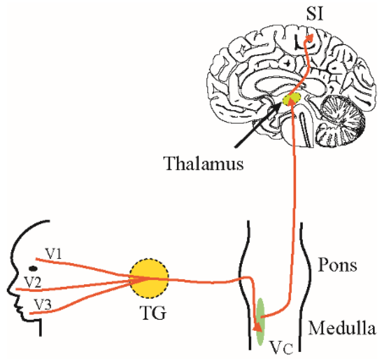
The trigeminal nerve is the biggest and most intricate of the 12 cranial nerves. It is the 5th and most developed cranial nerve with a wide distribution mechanism. This is a mixed nerve performing a gamut of motor somatic and sensitive fibers linked to the face. Primarily, the nerve is responsible for performing all sensitive innervation ... Trigeminal nerve. The large trigeminal nerve or 5th cranial nerve has three branches: ophthalmic (V1), maxillary (V2), and mandibular (V3) divisions. Trigeminal nerve is a mixed nerve providing sensations of the face for touch, temperature, and pain from the upper, middle, and lower portions of the face, as well as the oral cavity, to the brain. studies give information about the trigeminal and facial nerve. This paper will look at the anatomy of the trigeminal and facial nerves, common disorders of both nerves, specifics of NCS testing and case studies. Anatomy of the Trigeminal Nerve - For convenience, anatomy of the trigeminal nerve will be divided into three segments: brainstem,
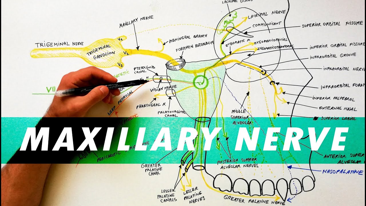
Trigeminal nerve branches diagram. The trigeminal nerve (the fifth cranial nerve, or simply CN V) is a nerve responsible for sensation in the face and motor functions such as biting and chewing; it is the most complex of the cranial nerves.Its name ("trigeminal" = tri-, or three, and - geminus, or twin: thrice-twinned) derives from each of the two nerves (one on each side of the pons) having three major branches: the ophthalmic ... Trigeminal Nerve. Create healthcare diagrams like this example called Trigeminal Nerve in minutes with SmartDraw. SmartDraw includes 1000s of professional healthcare and anatomy chart templates that you can modify and make your own. Symptoms. Trigeminal neuralgia results in pain occurring in an area of the face supplied by one or more of the three branches of the trigeminal nerve. Trigeminal neuralgia symptoms may include one or more of these patterns: Episodes of severe, shooting or jabbing pain that may feel like an electric shock. Spontaneous attacks of pain or attacks ... The maxillary nerve is the second branch of the trigeminal nerve, which originates embryologically from the first pharyngeal arch.. Its primary function is sensory supply to the mid-third of the face.. In this article, we shall look at the anatomy of the maxillary nerve - its anatomical course, sensory and parasympathetic functions.
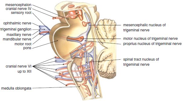
The mandibular nerve (third division of fifth cranial nerve, third division of trigeminal nerve, mandibular division of trigeminal nerve, CN V3, Latin: nervus mandibularis) is the third branch of the trigeminal nerve, a mixed nerve consisting of general somatic efferent (motor) and general somatic afferent (sensory) fibers.The sensory fibers of the mandibular nerve innervate several skin ... The trigeminal nerve roots. Both motor and sensory components of the trigeminal nerve complex exit the ventral mid-pons as distinct nerves.. The larger, more medial nerve is the trigeminal sensory root; and a smaller, more lateral nerve is the trigeminal motor root named portio minor (the minor portion of the trigeminal nerve; the fourth branch). These two nerve roots come together to form a ... Link to PayPal donation https://paypal.me/studentlamedicina?locale.x=en_US#anatomy #cranialnerve#maxillaryhttps://www.instagram.com/anatomy.knowledge/Anatomy... 2. Three nerve roots give rise to: a. Ophthalmic nerve, (CN V-1) b. Maxillary nerve, (CN V-2) c. Mandibular nerve, (CN V-3) 3. Peripheral distribution of three branches. Back of head and the angle of the jaw are not supplied by the trigeminal (Areas around ear supplied by CNs
Branches of the trigeminal nerve. Print. Sections. Products and services. Trigeminal neuralgia results in pain occurring in an area of the face supplied by one or more of the three branches of the trigeminal nerve. There is a problem with information submitted for this request. Review/update the information highlighted below and resubmit the form. This is a diagram of the trigeminal nerve, all three branches... some people are only affected in part of the nerve, one branch or even just part of a branch... I was "lucky" enough to have pain in all three branches as of this time. Find this Pin and more on Trigeminal Neuralgia info by Beth Elizabeth Worley. Trigeminal Neuralgia Treatment. The trigeminal nerve has three branches. It joins at the trigeminal ganglia and branches out to different parts of the face. Each branch division has a slightly different function. Start studying Trigeminal Nerve Distribution. Learn vocabulary, terms, and more with flashcards, games, and other study tools.
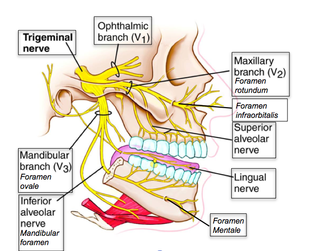
Trigeminal Nerve Innervation Diagram. Trigeminal Nerve Innervation Diagram. In this image, you will find ophthalmic branch, trigeminal nerve, maxillary brach, superior alveolar nerve, mandibular branch, inferior alveolar nerve, lingual nerve in it. Health care advices from Overseas Doctor . We are pleased to provide you with the picture named ...

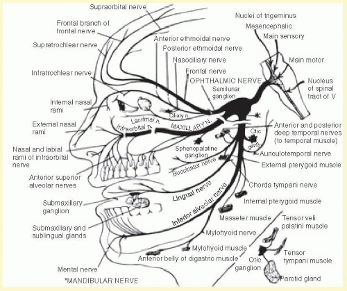
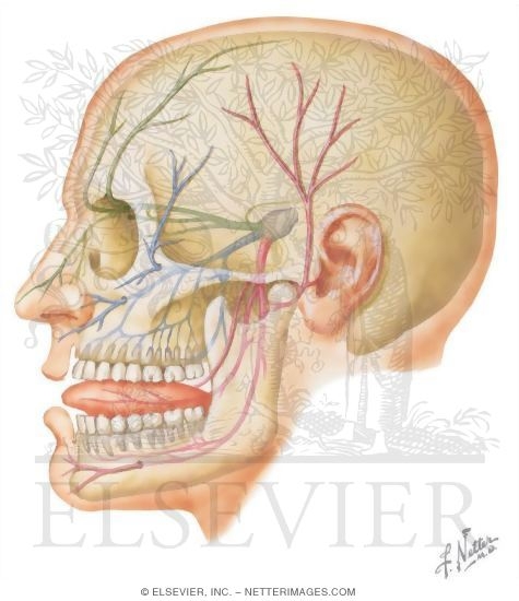
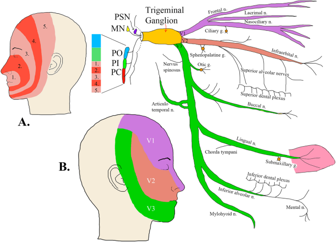
Diagram shows trigeminal nerve (TGN), trigeminal ganglion, and peripheral divisions and their branches. From fora-men rotundum ossis sphenoidalis, maxil-lary nerve (thin underline) gains access to pterygopalatine fossa and continues in floor of orbit as infraorbital nerve. Inferior alveolar and lingual nerves ( thick underline )
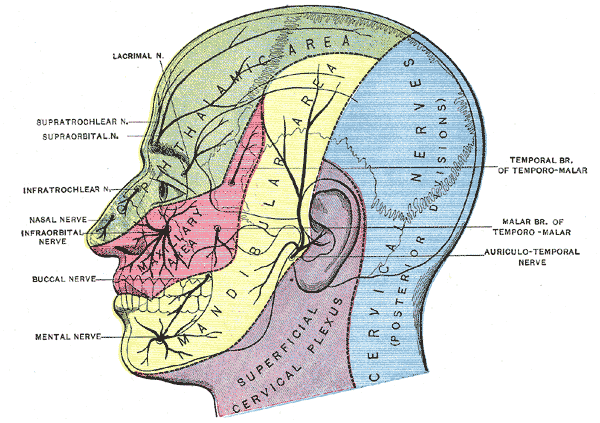
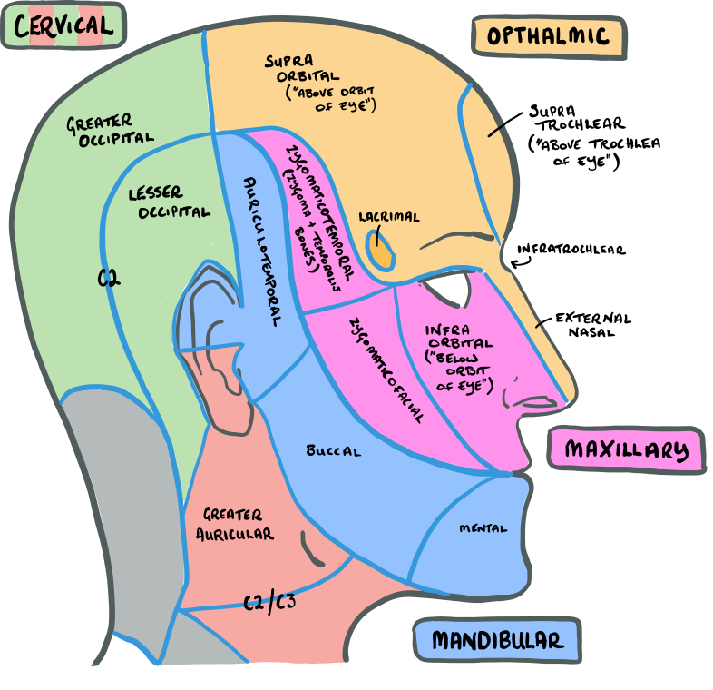
The trigeminal nerve is responsible for carrying most of the sensation of the face to the brain. The sensory trigeminal nerve branches of the trigeminal nerve are the ophthalmic, the maxillary, and the mandibular nerves, which correspond to sensation in the V1, V2, and V3 regions of the face, respectively. Ophthalmic nerve: This nerve detects ...

Trigeminal Nerve Anatomy: Gross Anatomy, Branches of the Trigeminal Nerve, Microscopic Anatomy The trigeminal nerve is the largest and most complex of the 12 cranial nerves (CNs). It supplies sensations to the face, mucous membranes, and other structures of the head.
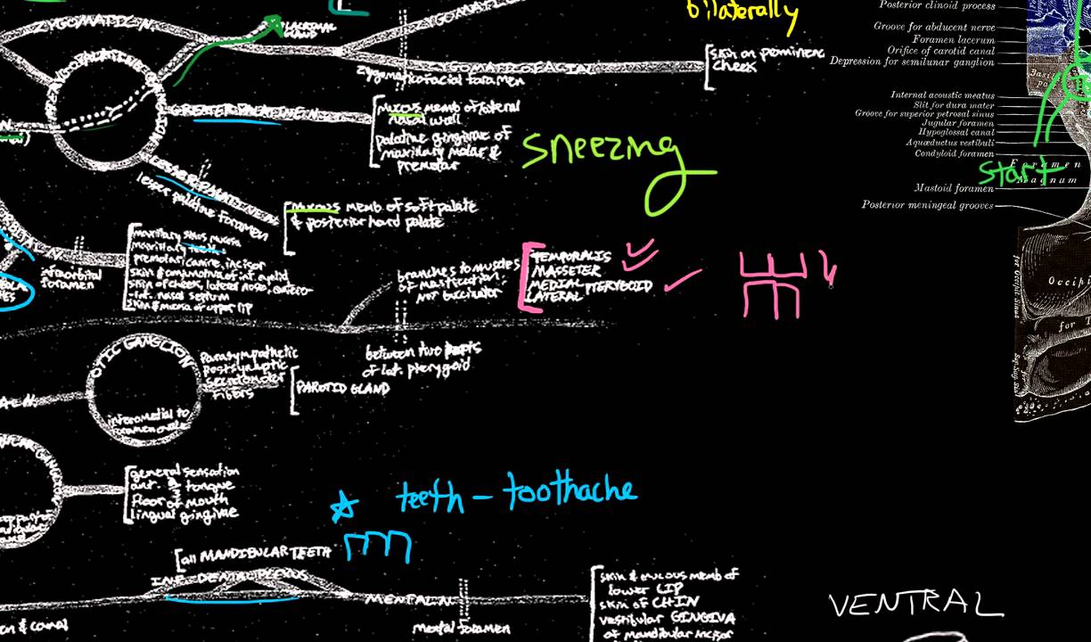
sensation includes: maxillary distal root of the 1st molar, maxillary 2nd and 3rd molars, periodontium and buccal gingiva, and part of the maxillary sinus. Mandibular division of the trigeminal nerve (V3) -mixed (motor and sensory) -largest of the 3 branches. -joins V2 at the trigeminal ganglion. -passageway through the foramen ovale.

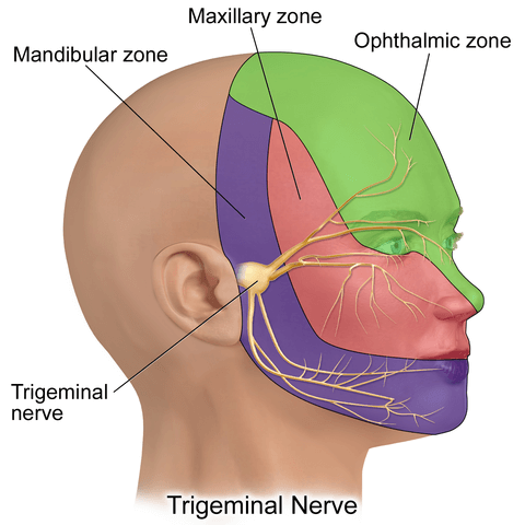
The trigeminal nerve contains three branches (that's why it's called trigeminal). The mandibular branch takes sensory data from the lower jaw area, all the way from the chin up to the area in front of the ear. The maxillary branch covers the area of the upper jaw, including the lower part of the nose and up to the area right below the eye.

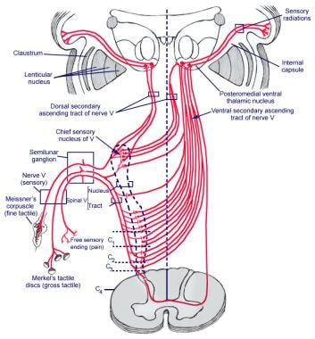
The trigeminal nerve is the largest cranial nerve and is the great sensory nerve of the head and face, and the motor nerve of the muscles of mastication.: It emerges from the side of the pons, near its upper border, by a small motor and a large sensory root—the former being situated in front of and medial to the latter.: Motor Root.—The fibers of the motor root arise from two nuclei, a ...
The trigeminal nerve is the fifth cranial nerve (CN V). Its primary function is to provide sensory and motor innervation to the face. The trigeminal nerve consists of three branches on either side that extend to different territories of the face. These branches join at the trigeminal ganglia which are located within the Meckel cave of the cranial cavity. The different branches are namely the ...
The trigeminal nerve is associated with derivatives of the 1st pharyngeal arch. Sensory: The three terminal branches of CN V innervate the skin, mucous membranes and sinuses of the face.Their distribution pattern is similar to the dermatome supply of spinal nerves (except there is little overlap in the supply of the divisions).

The trigeminal nerve, also known as the fifth (or V) cranial nerve, is a cranial nerve and its primary role is relaying sensory information from the face and head, although it does provide motor control to the muscles of mastication.It is both large and complicated and has multiple brainstem nuclei (sensory and motor) as well as many interconnections with other cranial nerves.

studies give information about the trigeminal and facial nerve. This paper will look at the anatomy of the trigeminal and facial nerves, common disorders of both nerves, specifics of NCS testing and case studies. Anatomy of the Trigeminal Nerve - For convenience, anatomy of the trigeminal nerve will be divided into three segments: brainstem,

Trigeminal nerve. The large trigeminal nerve or 5th cranial nerve has three branches: ophthalmic (V1), maxillary (V2), and mandibular (V3) divisions. Trigeminal nerve is a mixed nerve providing sensations of the face for touch, temperature, and pain from the upper, middle, and lower portions of the face, as well as the oral cavity, to the brain.

The trigeminal nerve is the biggest and most intricate of the 12 cranial nerves. It is the 5th and most developed cranial nerve with a wide distribution mechanism. This is a mixed nerve performing a gamut of motor somatic and sensitive fibers linked to the face. Primarily, the nerve is responsible for performing all sensitive innervation ...


0 Response to "36 trigeminal nerve branches diagram"
Post a Comment