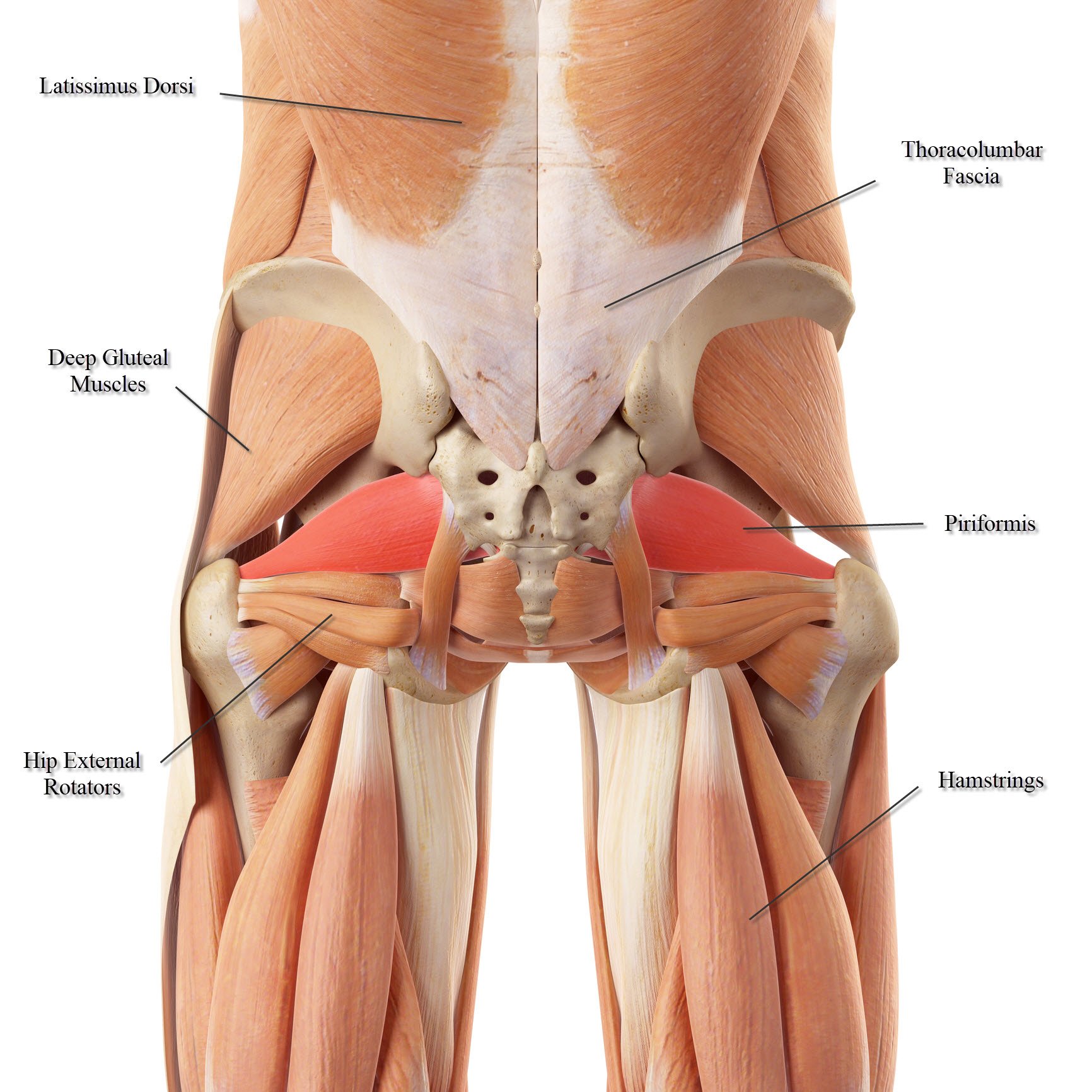
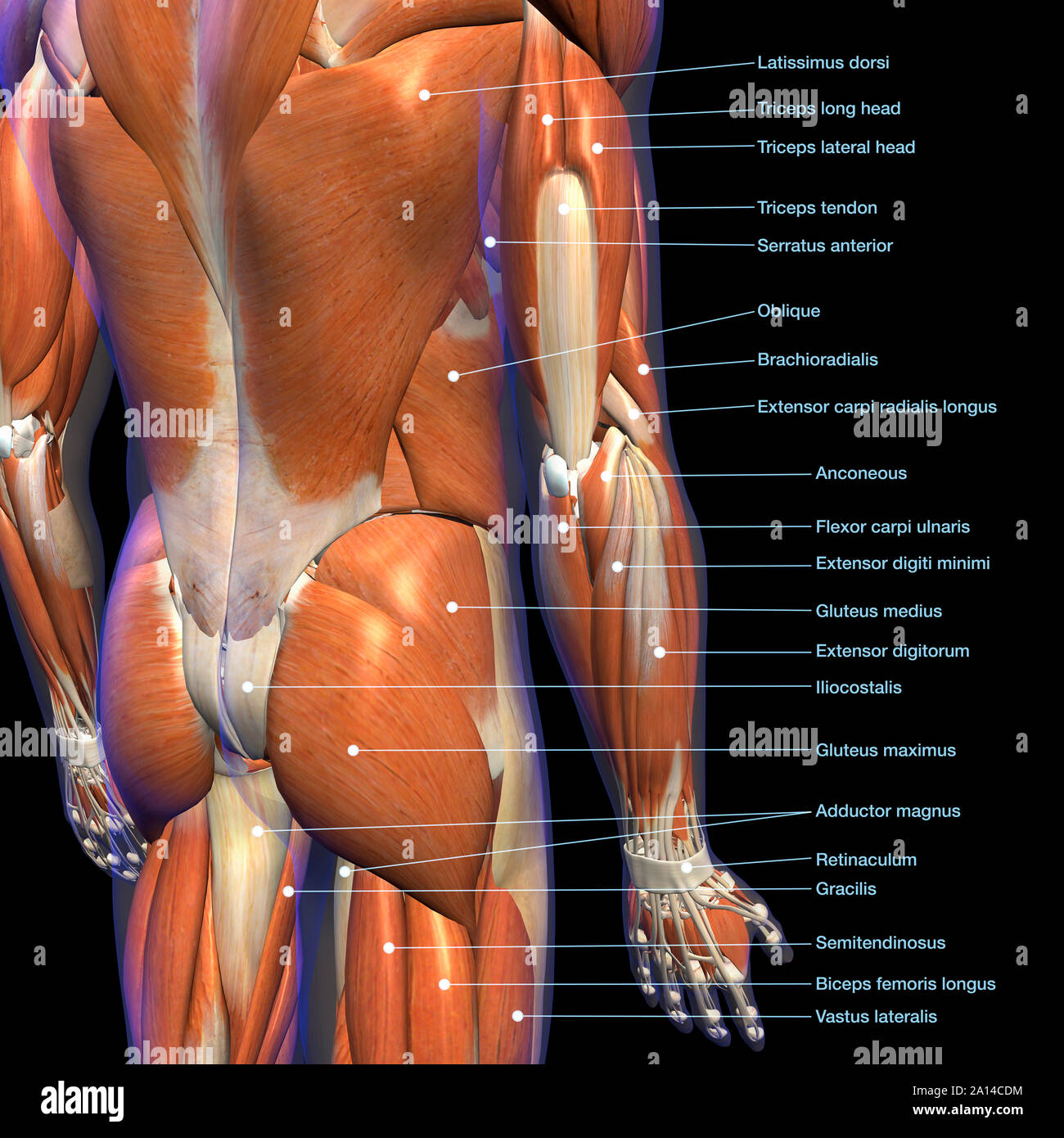
37 lower back muscle diagram human
This is a table of skeletal muscles of the human anatomy.. There are around 650 skeletal muscles within the typical human body. Almost every muscle constitutes one part of a pair of identical bilateral muscles, found on both sides, resulting in approximately 320 pairs of muscles, as presented in this article. Nevertheless, the exact number is difficult to define.
Are the pairs of protrusions on either side the vertebrae to which the back muscles attach. Illustration of the different sections of the spine. This diagram depicts blank muscle diagram 1024×659 with parts and labels. Human muscle system, the muscles of the human body that work the skeletal.
Muscle Anatomy Crossword - The Biology Corner. Muscle Anatomy Crossword. DOWN 1. helps regenerate ATP, ___ phosphate 3. thick filaments of a muscle fiber 5. type of muscle that connects to bones, voluntary 6. store neurotransmitters 7. neurotransmitter used to cause muscle contraction 9. connects muscles to bones 10. individual muscle fiber. 2.

Lower back muscle diagram human
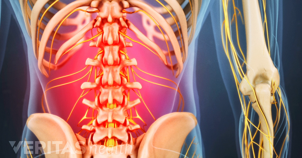
The part that forms the rear of any object or structure: Back definition, the rear part of the human body, extending from the neck to the lower end of the spine. The middle back consists of the spine (spinal column), spinal cord, nerves, discs, muscles, blood vessels, ligaments, and tendons. Low back pain is not a specific disease;
The larger, two-headed muscle on the back of your lower leg runs from the condyles (bony protrusions) on your thighbone to the Achilles tendon. Soleus. Originating below and beneath the gastrocnemius is the soleus muscle, which extends your foot when your knee is bent. Unlike the larger gastrocnemius, however, the soleus does not cross the knee ...
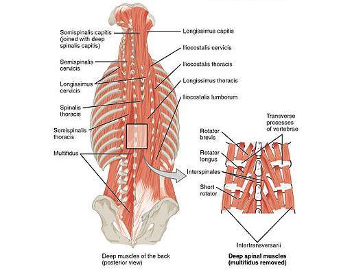
The muscles of the back categorize into three groups. The intrinsic or deep muscles are those muscles that fuse with the vertebral column. The second group is the superficial muscles, which help with shoulder and neck movements. The final group is the intermediate muscles, which help with the movement of the thoracic cage. Only the intrinsic muscles are considered true back muscles.
Lower back muscle diagram human.
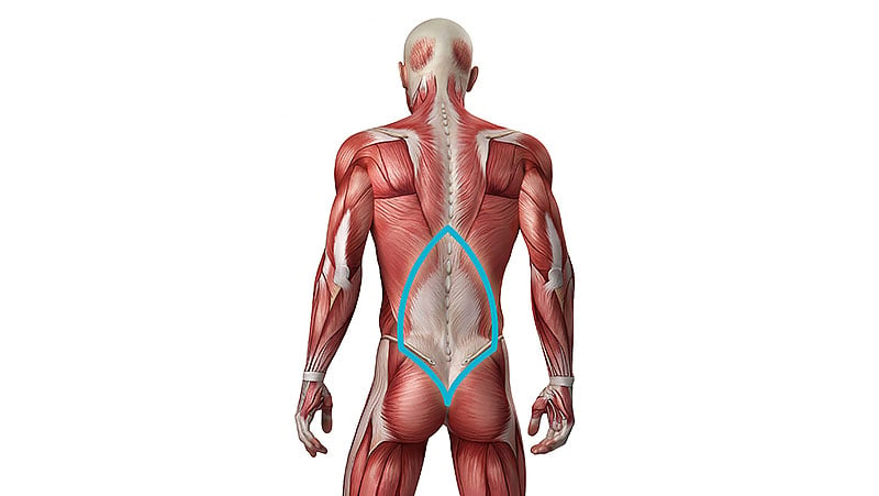
The muscles of the lower back help stabilize, rotate, flex, and extend the spinal column, which is a bony tower of 24 vertebrae that gives the body . · teres minor and infraspinatus: Be careful not to use the back muscles; Human muscle system, the muscles of the human body that work the skeletal.
Muscles are the only tissue in the body that has the ability to contract and therefore move the other parts of the body. Related to the function of movement is the muscular system's second function: the maintenance of posture and body position. Muscles often contract to hold the body still or in a particular position rather than to cause ...
Muscle Structure Diagram Back : Muscles Of The Lumbar Spine Of The Trunk /. The muscles of the lower back help stabilize, rotate, flex, and extend the. The occipitofrontalis muscle moves up the scalp and eyebrows. Feel the back of your ankle to. L2, l3, and l4 spinal nerves . Each of these parts are individual .
Human Anatomy Diagram Organs Back View : 386 Human Anatomy Organs Back View Photos And Premium High Res Pictures Getty Images. The spine provides support to hold the head and body up straight. The cornea, pupil, lens, iris, retina, and sclera. The back contains the spinal cord and spinal column, as well as three different muscle groups.
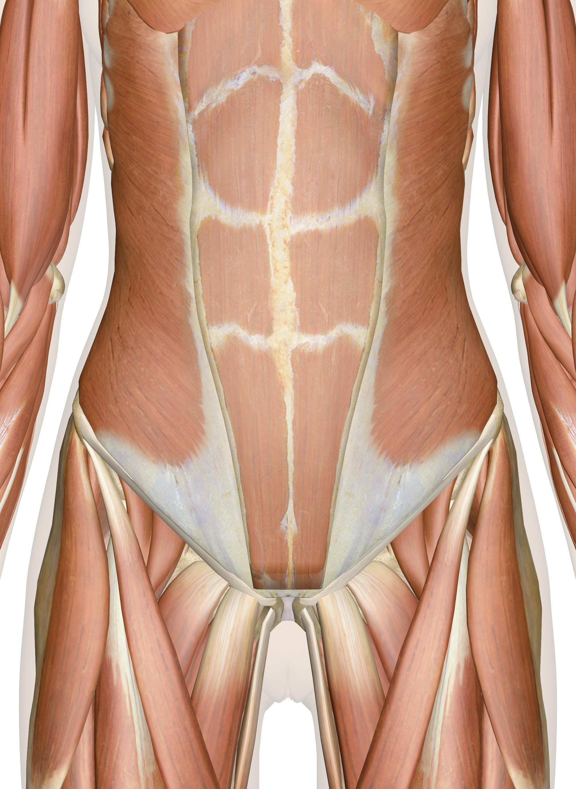
Superficial Skeletal Muscles. Most of the muscles shown move the skeleton for locomotion, but some muscles—especially those of the head—move other structures (e.g., the eyeballs, scalp, eyelids, skin of face, and tongue). The sheath of the left rectus abdominis, formed by aponeuroses of the flat abdominal muscles, has been removed to reveal ...
Lower limb (free PDF download) This muscle chart eBook covers the following regions: Inner hip & gluteal muscles. Anterior, medical and posterior thigh muscles. Anterior, lateral and posterior leg muscles. Dorsal and plantar foot muscles. This eBook contains high-quality illustrations and validated information about each muscle.
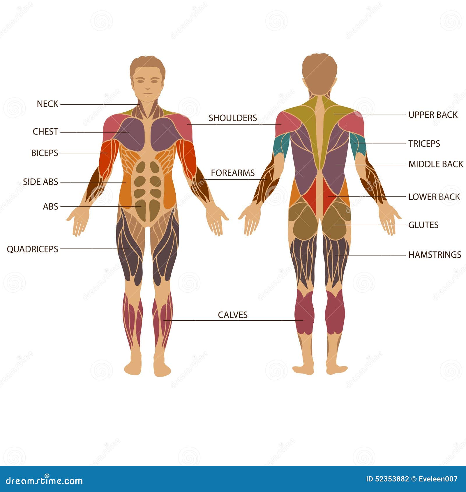
Human body muscle diagrams. Muscle diagrams are a great way to get an overview of all of the muscles within a body region. Studying these is an ideal first step before moving onto the more advanced practices of muscle labeling and quizzes. If you're looking for a speedy way to learn muscle anatomy, look no further than our anatomy crash courses .
Human Muscular Systems-Deep Layers of the Back Poster - Clinical Charts and Supplies from cdn11.bigcommerce.com This will help protect the muscles of your own back, shoulders, and arms. For your work area, such as a firm bed, padded floor, or massage table.
Related Posts of "Anatomy Of The Back Muscles" Abdominal Pictures Organs. Abdominal Pictures Organs 12 photos of the "Abdominal Pictures Organs" abdominal internal organs anatomy pictures, abdominal picture with organs, abdominal pictures organs, location of abdominal organs, picture abdominal organs female, Human Anatomy, abdominal internal organs anatomy pictures, abdominal picture with ...
Gluteus Medius. This muscle is a major generator of lower back and hip pain, as well as being responsible for complaints of a burning sensation along the posterior superior iliac spine (PSIS) and sacroiliac joint. Pain is often mistaken for lumbago- type pain, with discomfort (such as tenderness) into the buttocks and superior thigh.
Aug 19, 2016 - Bone And Lower Back Muscles Lower Back Muscles Anatomy Human Anatomy Diagram.
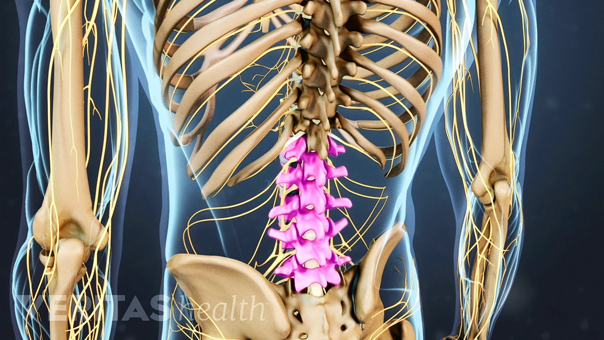
740 lumbar spine anatomy diagram stock photos, vectors, and illustrations are available royalty-free. See lumbar spine anatomy diagram stock video clips. of 8. spinal vertebrae bone spine vertebra toracica spinal cord spine structure back diagram spine sections spinal cord vertebrae spinal structure health diagram. Try these curated collections.
The muscles of the lower back help stabilize, rotate, flex, and extend the spinal column, which is a bony tower of 24 vertebrae that gives the body ...
Anatomical diagrams of the spine and back. This human anatomy module is composed of diagrams, illustrations and 3D views of the back, cervical, thoracic and lumbar spinal areas as well as the various vertebrae. It contains the osteology, arthrology and myology of the spine and back. It is particularly interesting for physiotherapists ...
Extensor, Flexor and Oblique Muscles and Back Pain · The extensor muscles are attached to back of the spine and enable standing and lifting objects. · The flexor ...
52,129 human back anatomy stock photos, vectors, and illustrations are available royalty-free. See human back anatomy stock video clips. of 522. human body anatomy female female anatomy muscle shoulder blade pain anatomy back muscles bones man female anatomy body muscles in a body female anatomy muscole shoulder concept muscular sysyem.
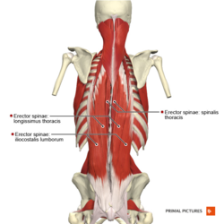
They are the spinalis, iliocostalis, and longissimus. Each of these muscles is divided into three parts that correspond to the part of the back where they're ...
Back Muscle Chart. Human Muscles · December 13, 2020. Back Muscle Chart.
The lumbar and sacrum region make up the bone of the lower back anatomy. The spinal cord is contained within the spine's vertebrae, running through the ...






:background_color(FFFFFF):format(jpeg)/images/article/en/intrinsic-back-muscles/gyxZt86ah5Dh2cGeXpQ_Deep_back_muscles.png)
:background_color(FFFFFF):format(jpeg)/images/library/12060/Img.jpg)


:background_color(FFFFFF):format(jpeg)/images/library/12644/Ventral_trunk_muscles.png)



:background_color(FFFFFF):format(jpeg)/images/library/14074/Back_muscles.png)




0 Response to "37 lower back muscle diagram human"
Post a Comment