40 diagram of sinus cavity
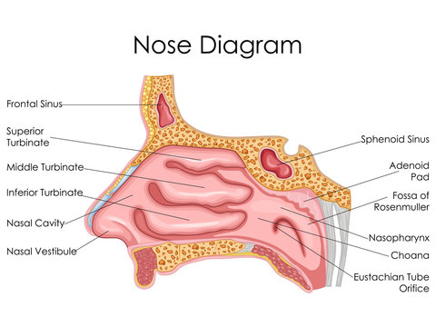
SINUS CAVITIES DIAGRAM by Sinus Help. www.sinusinfectionrelief.com. Fundamentally situated in the skull, human sinus cavities are the passageways mainly found in the areas around the face. The nose and the sinuses are closely linked by the ostium. The nose is divided into two cavities by the nasal septum. The superior, middle and inferior ...
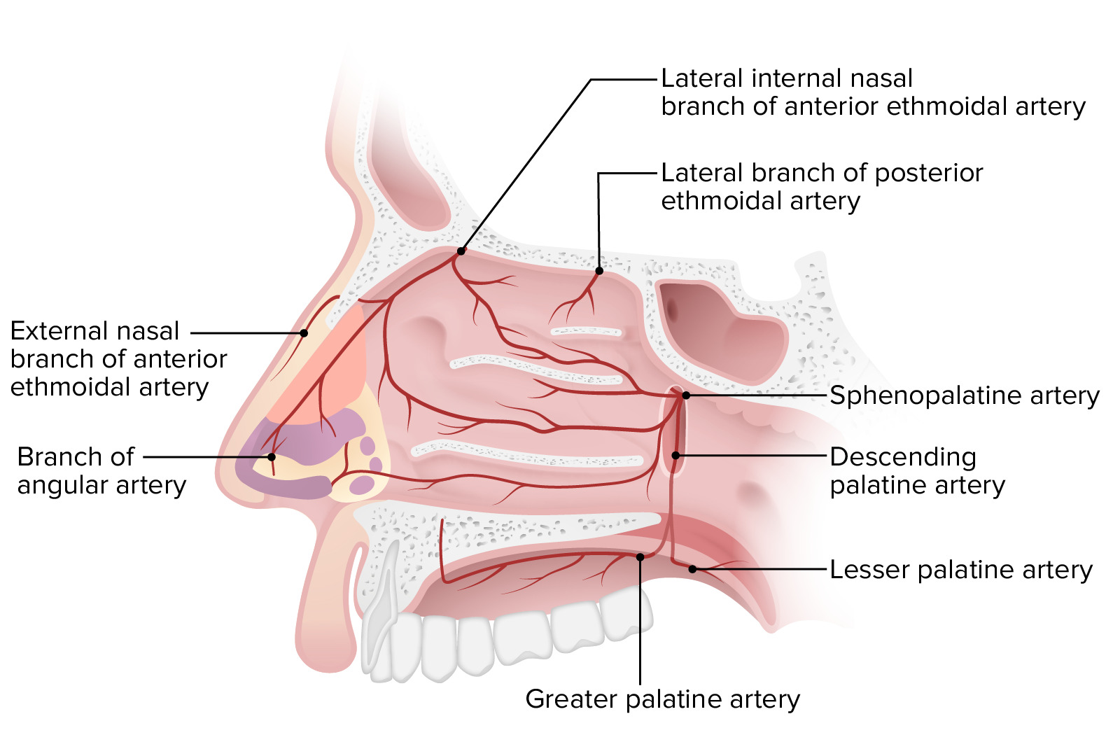
Nose. The external nasal anatomy is quite simple. It is a pyramidal structure, with its root located superiorly and apex sitting inferiorly.The root is continuous with the anterior surface of the head and the part between the root and the apex is called the dorsum of the nose. Inferior to the apex are the two nares (nostrils), which are the openings to the nasal cavity.

Diagram of sinus cavity
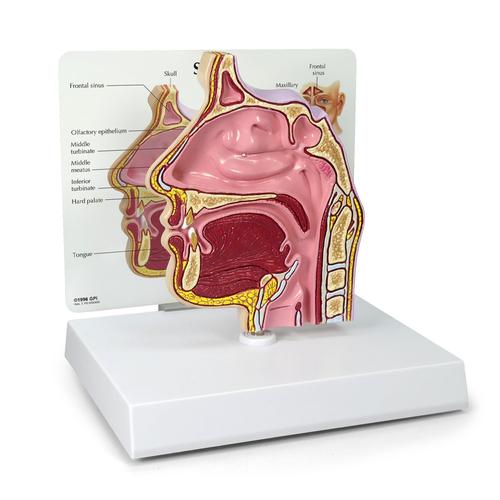
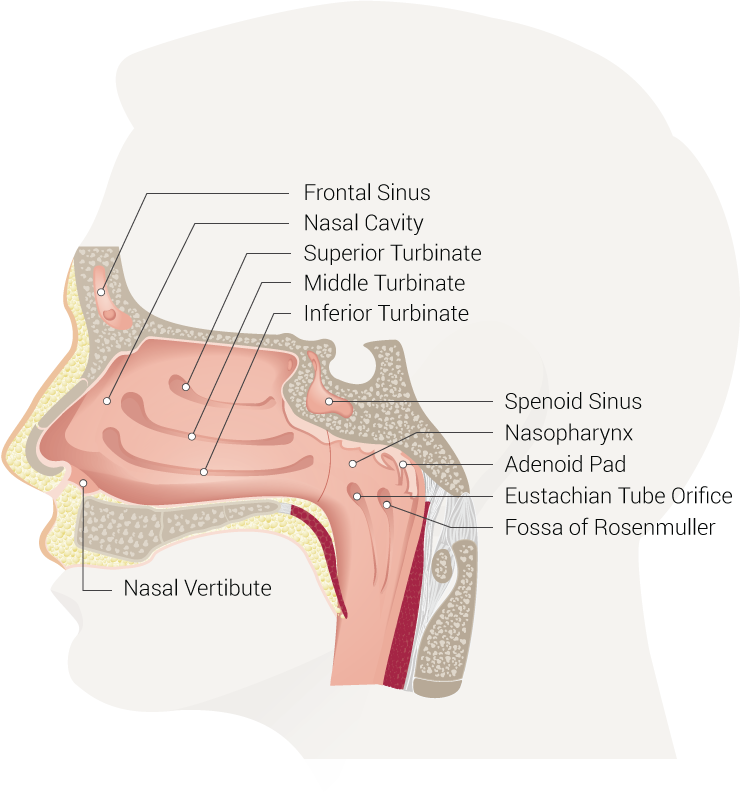
Like the nasal cavity, the sinuses are all lined with mucus. The mucus secretions produced in the sinuses are continually being swept into the nose by the hair-like structures on the surface of ... Anatomy of the nasal mucosa. The nasal mucosa, also called respiratory mucosa, lines the entire nasal cavity, from the nostrils (the external openings of the respiratory system) to the pharynx (the uppermost section of the throat).The external skin of the nose connects to the nasal mucosa in the nasal vestibule. A dynamic layer of mucus overlies the nasal epithelium (the outermost layer of ... The nose is an olfactory and respiratory organ. It consists of nasal skeleton, which houses the nasal cavity. The nasal cavity has four functions: Warms and humidifies the inspired air.; Removes and traps pathogens and particulate matter from the inspired air. Responsible for sense of smell. Drains and clears the paranasal sinuses and lacrimal ducts.
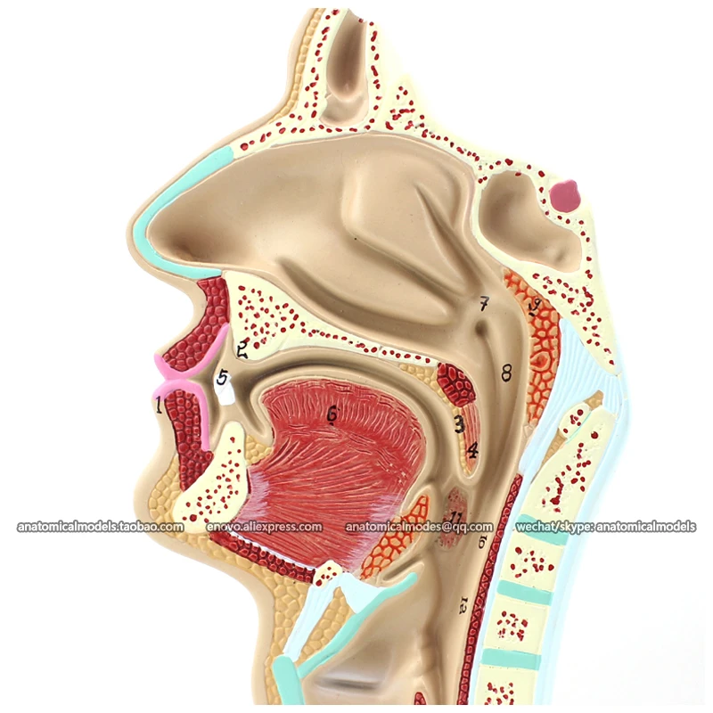
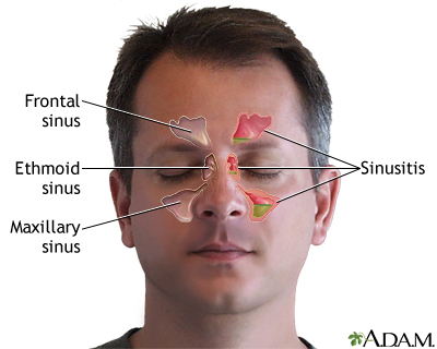
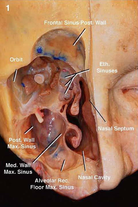
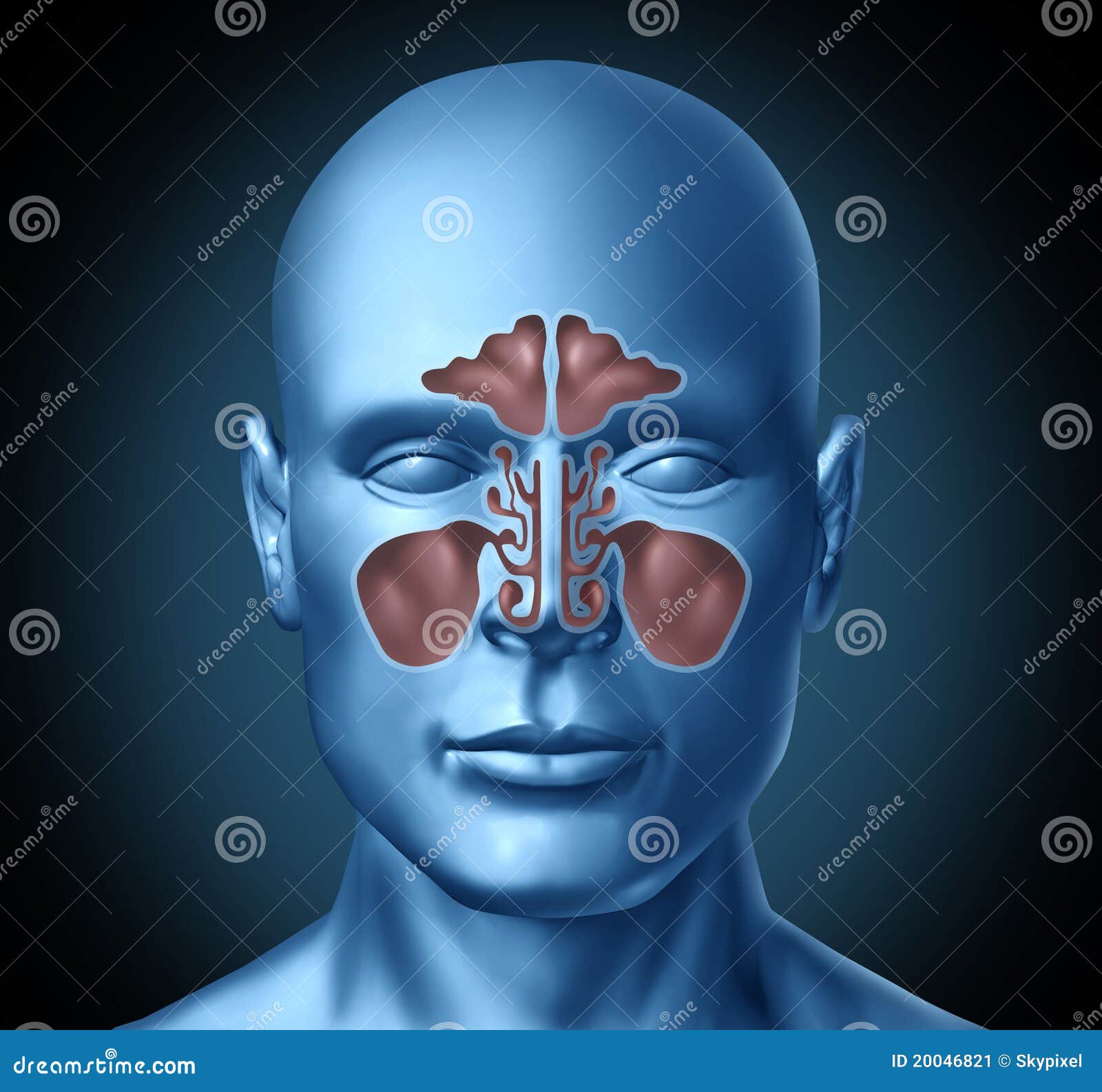
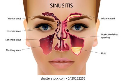
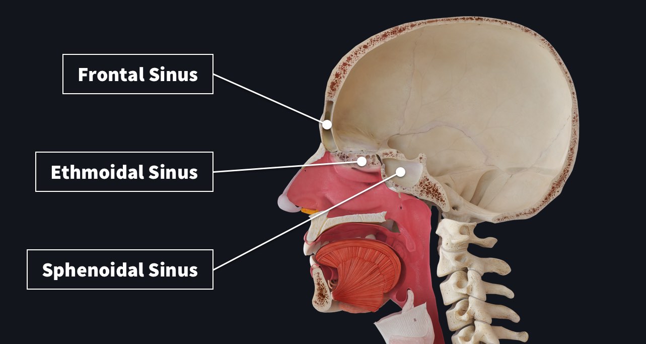
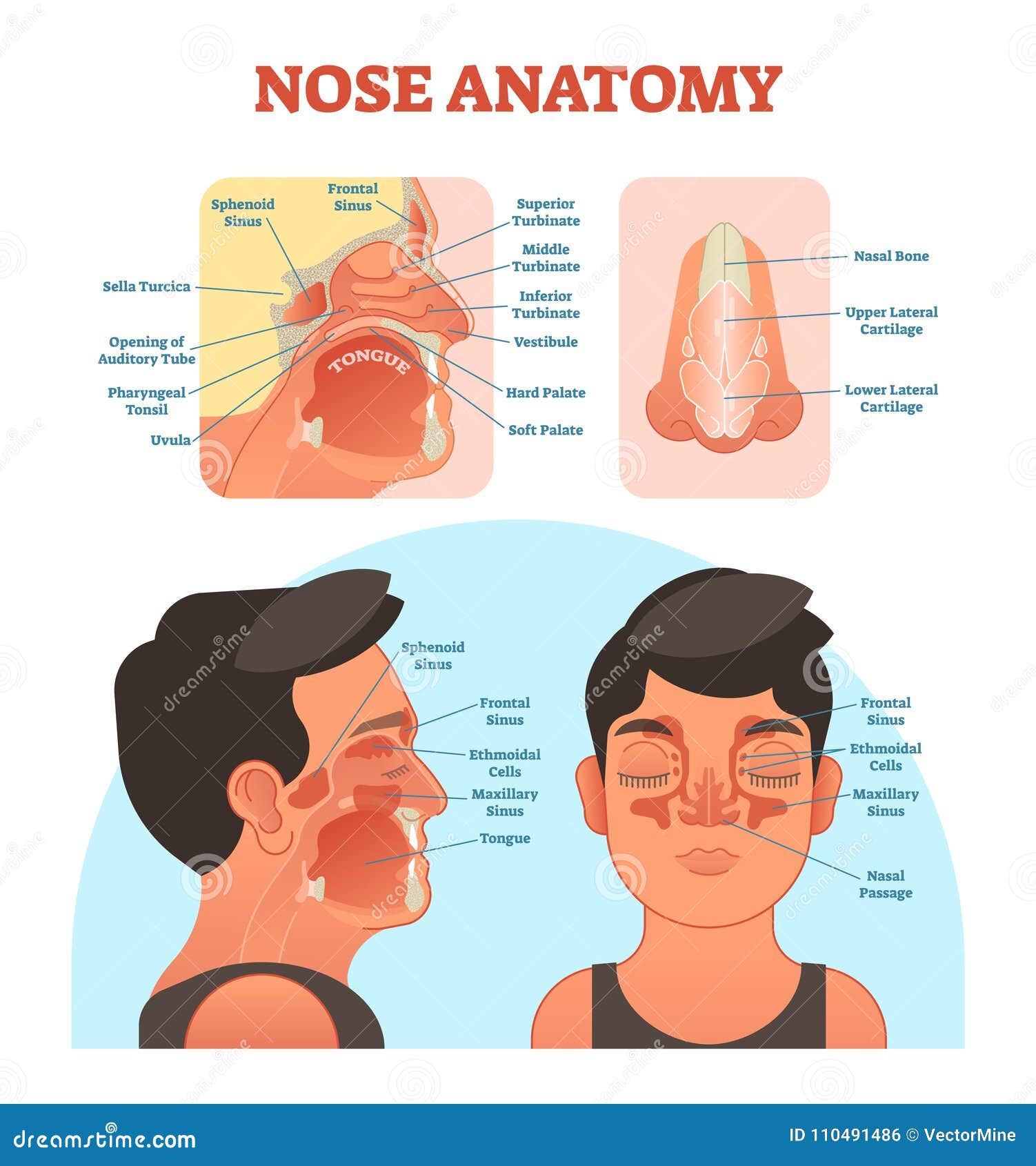
Diagram of sinus cavity. The nasal cavity is a roughly cylindrical, midline airway passage that extends from the nasal ala anteriorly to the choana posteriorly.[1] It is divided in the midline by the nasal septum. On each side, it is flanked by the maxillary sinuses and roofed by the frontal, ethmoid, and sphenoid sinuses in an anterior to posterior fashion.[1] While seemingly simple, sinonasal anatomy is composed of ... The cavernous sinuses are 1 cm wide cavities that extend a distance of 2 cm from the most posterior aspect of the orbit to the petrous part of the temporal bone.They are bilaterally paired collections of venous plexuses that sit on either side of the sphenoid bone.Although they are not truly trabeculated cavities like the corpora cavernosa of the penis, the numerous plexuses, however, give the ... The paranasal sinuses are air-filled extensions of the nasal cavity. There are four paired sinuses - named according to the bone in which they are located - maxillary, frontal, sphenoid and ethmoid. Each sinus is lined by a ciliated pseudostratified epithelium, interspersed with mucus-secreting goblet cells. Diagram of Sinus Cavities and Drainage Ports What is Sinusitis? Sinusitis is caused by an inflammation of your sinus cavities that causes redness, swelling, mucus, and pain. There are two types of sinusitis: Acute sinusitis - an infection that is often triggered by the flu or cold. The flu or cold virus attacks your sinuses causing them to ...
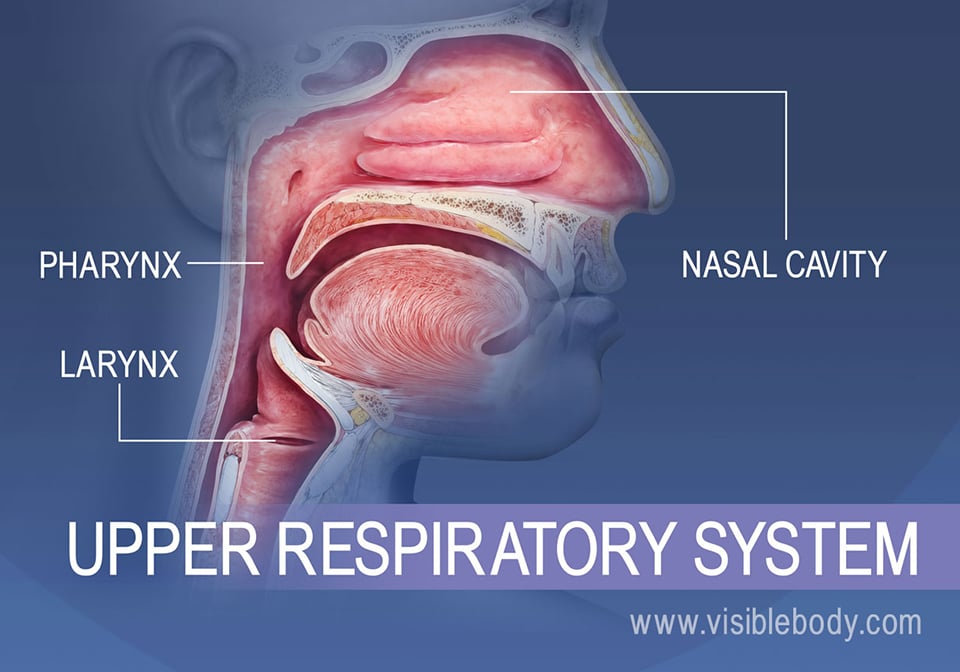
Nasal Cavity Definition. The nose is one of the primary sensory organs responsible for the sense of smell, while it also plays major roles in respiration and speech production [1].The nasal cavity lies just behind the two nostrils and forms the interiors of the nose.. It makes up the upper respiratory system along with the paranasal sinuses, oral cavity, pharynx, and larynx [2], and is the ... Sinus Cavities Diagram. 1. SINUS CAVITIES DIAGRAM by Sinus Help www.sinusinfectionrelief.com. 2. Fundamentally situated in the skull, human sinus cavities are the passageways mainly found in the areas around the face. Also known as paranasal sinuses, they are hollow, irregular air cavities which sit adjacent to and are attached to the nose and ... The rest of the sinus cavity is empty and filled with air. Frontal Sinus. These are two large sinuses located on either side of the anterior part of the frontal bone (the front of the frontal bone) just above the orbits. The left and right frontal sinuses which usually differ in size have two parts - a horizontal and vertical part. Sinuses are open cavities in the head, but they are not bare bones. Each sinus cavity has a mucous membrane lining that allows very slow ventilation into and out of the sinus. The slow ventilation allows the space in sinus cavities to be filled with a high concentration of carbon dioxide and a low level of oxygen.
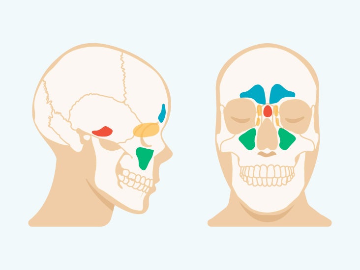
Title: how to draw nasal cavity | how to draw nasal cavity easy | how to draw nasal cavity step by step@AsapKNOWLEDGE Hello Friends in this video I tell you... There are four pairs of sinuses (named for the skull bones in which they're located). Interactive diagrams show sinus cavity locations and help visualize ...Sinus diagram · Function · Sinusitis · Sinusitis picture The maxillary sinuses are the pockets near the cheeks. This sinus is located in the maxilla bone under the eye, contains three recesses, and is shaped like a pyramid. The frontal sinuses are over the forehead and above the eyes. These are absent at birth, and in approximately 5% of the adult population. The ethmoid sinuses are actually several ... View nasal cavity diagram videos. Browse 35 nasal cavity diagram stock illustrations and vector graphics available royalty-free, or start a new search to explore more great stock images and vector art. antique illustration: head throat section - nasal cavity diagram stock illustrations. cutaway diagram of human respiratory system, including ...
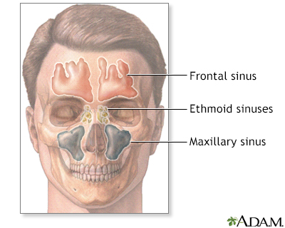
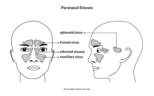
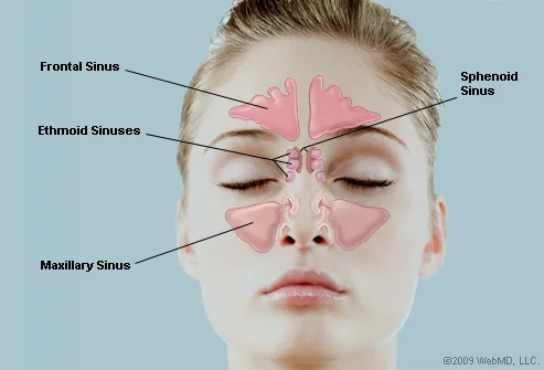
Sinuses are hollow, air-filled cavities that are located throughout the skull. There are several sets of sinuses in the head. The maxillary sinuses are behind the cheeks. The ethmoid sinuses lie under the inside corners of the eyes. The sphenoid sinuses are located behind the ethmoid sinuses. The frontal sinuses lie behind the forehead above ...
Sinus Pressure Points for Sinus Relief. Below are a number of sinus trigger points to help alleviate the discomfort from sinus pressure; some will even help mucus drain. A sinus pressure points diagram is also included to assist you in finding the location accurately on your face:
The sinuses are a connected system of hollow cavities in the skull. The largest sinus cavities are about an inch across. Others are much smaller. Your cheekbones hold your maxillary sinuses (the ...
Browse 37 nasal cavity diagram stock photos and images available, or start a new search to explore more stock photos and images. antique illustration: head throat section - nasal cavity diagram stock illustrations. cutaway diagram of human respiratory system, including nasal and mouth cross section. - nasal cavity diagram stock illustrations.
The sinuses are an air-filled cavity in a dense portion of a skull bone. They actually decrease the weight of the skull. The sinuses are formed in four right-left pairs. The frontal sinuses are positioned behind the forehead, while the maxillary sinuses are behind the cheeks. The sphenoid and ethmoid sinuses are deeper in the skull behind the ...
A sinus is a sac or cavity in any organ or tissue, or an abnormal cavity or passage caused by the destruction of tissue.In common usage, "sinus" usually refers to the paranasal sinuses, which are air cavities in the cranial bones, especially those near the nose and connecting to it. Most individuals have four paired cavities located in the cranial bone or skull.
by B Henson · 2020 · Cited by 2 — The word “sinus” is most commonly understood to be the paranasal sinuses that are located near the nose and connect to the nasal cavity.
Maxillary Sinus (within the maxillary bones): The largest among all the paranasal sinuses [2], these two conical cavities are located on the two sides of the nose, above the upper teeth, and below the cheeks [4]. Ethmoid Sinus (within the ethmoid bones): Three to eighteen [5] air cells present in the ethmoid labyrinth, on both sides of the nose, between the eyes [6, 7].
The entire system is surrounded by a system of hollow cavities in the skull called the sinuses. The sinuses are about an inch across and are located in the cheekbones, the center of the forehead, between the eyes and in the nose. This interconnected ENT system helps us to breathe, smell and taste and plays a defining role in our looks.
Where do I get my information from: http://armandoh.org/resourceFacebook:https://www.facebook.com/ArmandoHasudunganSupport me: http://www.patreon.com/armando...
Sinuses of Nose. Human Anatomy - Sinus Diagram. Anatomy of the Nose. Nasal cavity bones. Anatomy of paranasal sinuses. Sinusitis - It is the inflammation of
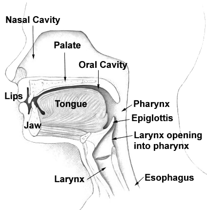
The nasal cavity consists of all the bones, tissues, blood vessels and nerves that make up the interior portion of the nose. The most important functions of the nasal cavity include warming and humidifying the air as you breathe and acting as a barrier for the immune system to keep harmful microbes from entering the body.
The nose is an olfactory and respiratory organ. It consists of nasal skeleton, which houses the nasal cavity. The nasal cavity has four functions: Warms and humidifies the inspired air.; Removes and traps pathogens and particulate matter from the inspired air. Responsible for sense of smell. Drains and clears the paranasal sinuses and lacrimal ducts.
Anatomy of the nasal mucosa. The nasal mucosa, also called respiratory mucosa, lines the entire nasal cavity, from the nostrils (the external openings of the respiratory system) to the pharynx (the uppermost section of the throat).The external skin of the nose connects to the nasal mucosa in the nasal vestibule. A dynamic layer of mucus overlies the nasal epithelium (the outermost layer of ...
Like the nasal cavity, the sinuses are all lined with mucus. The mucus secretions produced in the sinuses are continually being swept into the nose by the hair-like structures on the surface of ...






(166).jpg)






0 Response to "40 diagram of sinus cavity"
Post a Comment