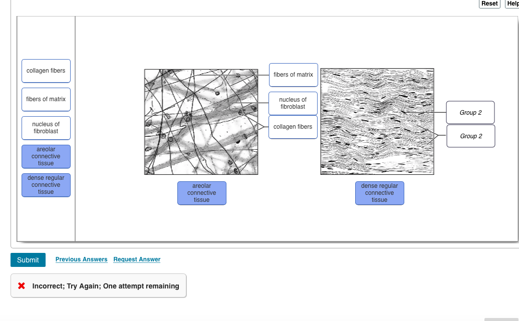
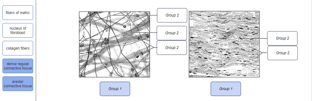
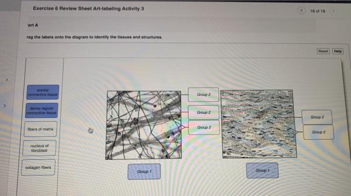
40 drag the labels onto the diagram to identify the types of connective tissue proper.
Password requirements: 6 to 30 characters long; ASCII characters only (characters found on a standard US keyboard); must contain at least 4 different symbols;
Epimysium. connective tissue layer covering the outermost part of skeletal muscle. Protects the muscle from damage by friction, carries blood vessels & nerves to the muscle, and helps to form the tendon. Perimysium. Drag the labels onto the diagram to identify structural features associated with skeletal muscle.
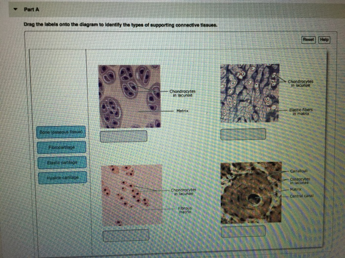
Drag the labels onto the diagram to identify the structures found in compact bone. It looks like a sponge or honeycomb with a lot of spaces in between. When the posterior structures of the glenohumeral joint are shortened, this in fact, some authors have identified internal impingement as the leading cause of rotator cuff.

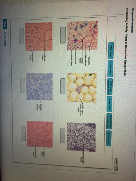
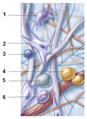
Drag the labels onto the diagram to identify the types of connective tissue proper.
Developing Data Flow Diagrams 31 Introduction 32 Context diagram 33 Level 1 Data Flow Diagram 34 Lower levels of Data Flow Diagrams 35 Check list 4. From the Diagram Toolbar drag Process onto the diagram. A Data Flow diagram DFD is a graphical representation of the flow of data through an information system.
What is Areolar Connective Tissue? Areolar connective tissue is a loosely arranged connective tissue that is widely distributed in the body and contains collagen fibers, reticular fibers, and a few elastic fibers embedded in a thin, almost fluid-like ground substance. The areolar connective tissue is a subtype of loose connective tissue.. It is the commonest type of connective tissue in the ...
Drag the labels onto the diagram to identify the various types of cutaneous receptors. Reset Help G Free nerve endings (pain temperature) Lamellar corpuscle (deep pressure) Dermis Tactile corpuscle (touch, light pressure) Epidermis Hair follicle receptor (hair movement, light touch) Bulbous corpuscle (deep continuous pressure)
Drag the labels onto the diagram to identify the types of connective tissue proper..
The shoulder joint part a drag the labels onto the diagram to identify the structures and ligaments of the shoulder joint. Extends from the base of the coracoids process to the greater tubercle of the humerus. This diagram here just shows the joint capsule itself.
Smooth Muscle Diagram Labeled - Draw Well Labelled Diagram Of Various Types Of Muscle s Present In Human Body Brainly In ... Smooth muscle is composed of sheets or strands of smooth muscle cell s. It is the pen diagram of skeletal, smooth and cardiac muscle for class 10, 11 and 12. Give one difference between yudem and nh. Structure. Smooth muscle cell s are pivot-shaped; hold one centrally ...
Academia.edu is a platform for academics to share research papers.
2,459 Likes, 121 Comments - University of South Carolina (@uofsc) on Instagram: “Do you know a future Gamecock thinking about #GoingGarnet? 🎉 ••• Tag them to make sure they apply…”
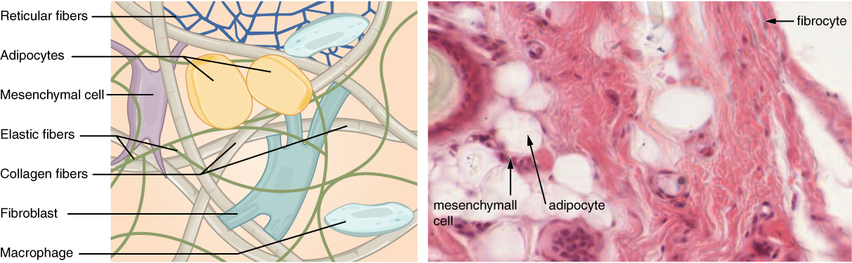
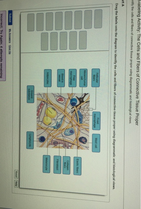
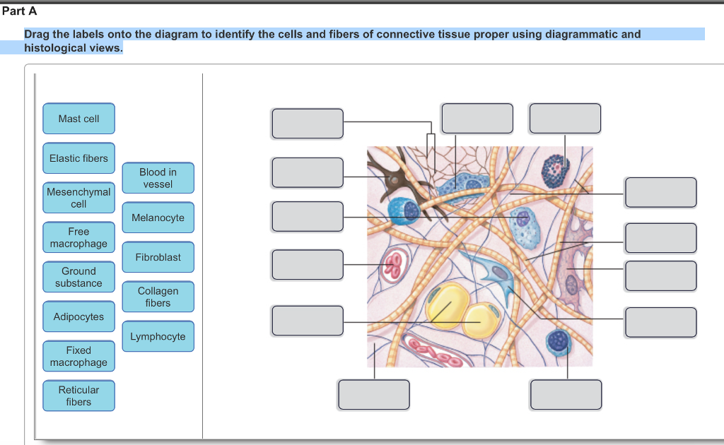
Drag the labels onto the diagram to identify the cells and fibers of connective tissue proper using diagrammatic and histological views. Image: Reticular Fibers ...
The primary function of the kidney is to extract water accompanied with other constituents from the blood. Unlabelled diagram of the muscles of the body for pupils to complete. First, we have the identifying proper names for each muscle. Muscle diagrams are a great way to get an overview of all of the muscles within a body region.
Feb 11, 2020 · @alwaysclau: “It’s quite an experience hearing the sound of your voice carrying out to a over 100 first year…”
Treatment. The trachea, commonly known as the windpipe, is the large tube that delivers air from the upper respiratory tract (the nasal passages, throat, and larynx) to the bronchi (the two large airways that branch off into each lung). In the process, it warms and moisturizes the air and catches debris and microbes before they enter the lungs. 1.
Drag the labels onto the diagram to identify the parts of the placenta and ... Identify the connective tissue proper cellular component labeled "F".
Page 3. Labelled diagram of Bones of the face. Page 4. Labelled diagram of the bones neck, chest & shoulder. Task 2 Page 5. Labelled diagram showing the position of the Muscles of the face. Page 6. Labelled diagram showing the Muscles that move the head Page 7 .Chart showing the action and location of the muscle of the face. Task 3 Page 8 (A).
The Renin-Angiotensin-Aldosterone System (RAAS) is a hormone system within the body that is essential for regulation of blood pressure and fluid balance. It is comprised of the three hormones renin, angiotensin II and aldosterone and regulated primarily by renal blood flow. This article shall discuss the system, how it is regulated and clinically relevant conditions to its dysfunction.
Drag the labels onto the diagram to identify the different types of gated ion channels. The concentrations of which two ions are highest outside the cell? - Na+ and protein anions (A−) - K+ and protein anions (A−) - Na+ and Cl− - K+ and Cl−. Na+ and Cl−. Let's consider a scenario in which the resting membrane potential changes from −−70 mVmV to +70 mVmV, but the …
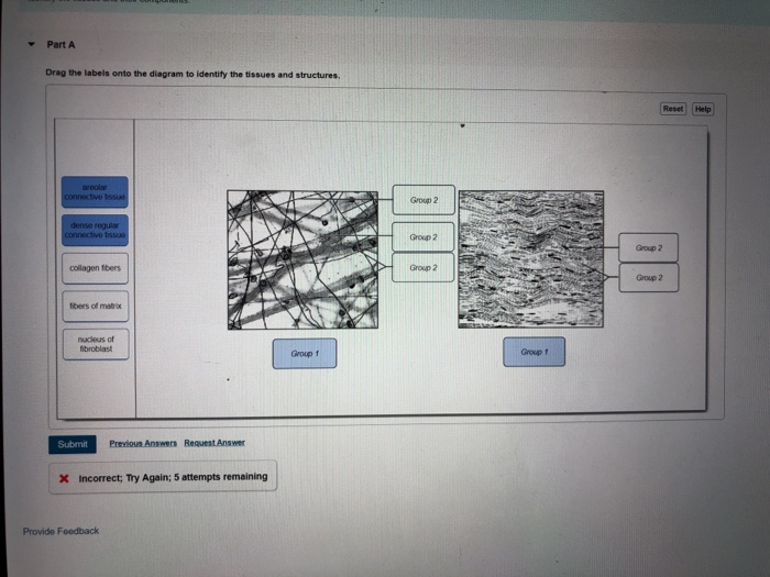
Drag the labels onto the diagram to identify the tissues and ...
Epithelial tissue covers the cavities and surfaces of the body's organs. Learn about the types, function, and structure of epithelial tissue, including pseudostratified columnar epithelium and ...

David w grainger's research works | university of utah, utah (uou ...
Anatomy Structure . Within the gluteus maximus, fibers from the muscle enter into different parts of the body. This includes the femur (also known as the thighbone) and the iliotibial tract or band, which is made up of connective tissue that runs up the thigh. The area of the gluteus maximus known as the gluteal crease (also called the gluteal sulcus) is known as the horizontal crease right ...
Solved drag the labels onto the diagram to identify the | chegg.com
Kidney Structures and Functions Explained (with Picture and Video) Your kidneys are paired organs found on each side of the back portion of the abdominal cavity. The larger left kidney is located a bit higher than the right kidney. Unlike other organs found in the abdomen, the kidneys are located behind the lining (peritoneum) of the abdominal ...

Solved drag the labels onto the diagram to identify the | chegg.com
Drag the labels onto the diagram to identify the various types of ... Which component of the connective tissue in this field of view is highlighted? Rating: 5 · 1 review
Solved -aibrocytes reticular fbers bundles part a drag the | chegg.com
40 drag the labels onto the diagram to identify the types of epi the lia. Written By Kathleen D. Walker Sunday, December 5, ... Drag the labels onto the diagram to identify the divisions and recep to rs of the nervous system. look at pic Drag the labels to identify the structural components of a typical neuron.

Taste bud - wikipedia
which type of muscle tissue is under your conscious control? skeletal muscle tissue. what cell is activated during an injury to connective tissue that ...

Ch 13 lab map, ch 12 lab map, ch 11 lab map, ch 10 lab map, ch 13 ...
Correctly label the following parts of a mucus membrane. Determine which connective tissue type each image below represents. Then click and drag the labels matching them up with the correct tissue type. Click and drag each label into the appropriate category according to which type of epithelia it pertains.

Departemen histologi dan patologi anatomi, fakultas kedokteran ...
Drag the labels onto the diagram to identify the divisions and receptors of the nervous system. look at pic Drag the labels to identify the structural components of a typical neuron. Transcribed image text: Drag the labels onto the diagram to identify the divisions and receptors of the nervous system. Reset Help Organization of the Nervous System Integrate, process, and coordinate sensory data ...

Drag the labels onto the diagram to identify the structures and ...
Question: Drag the labels onto the diagram to identify structural features of epi the lium. Connective tissue Basement membrane Nucleus IDIO Lumen of duct S ... Transcribed image text: Art-labeling Activity: Classifying Epithelia Drag the labels onto the diagram to identify the types of epithelia. Color breathing is a simple stress reducing activity that may be quickly learned.
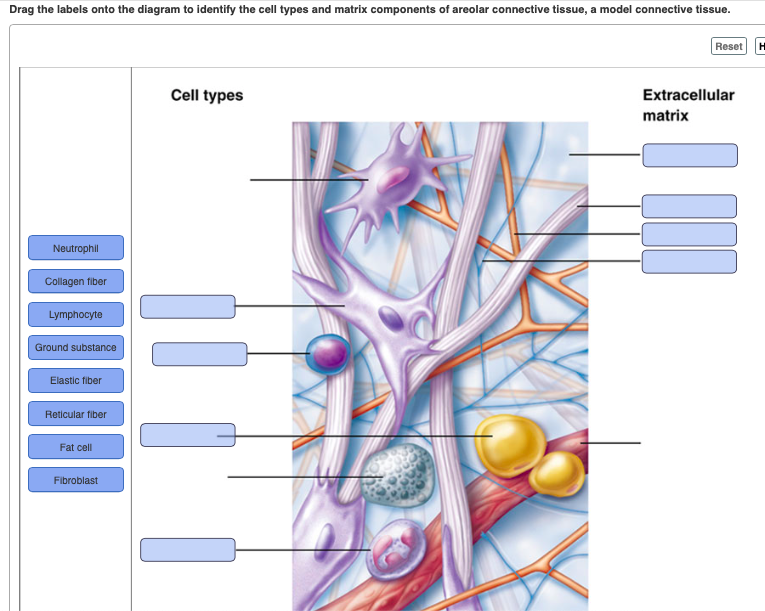
Solved drag the labels onto the diagram to identify the cell ...
Drag the labels to their appropriate targets to correctly identify the various chromosome structures. Determine whether each of the following describes the male or female skeleton. The structure of bone tissue suits the function. Drag each label into the proper position in order to identify the effect of each condition on blood calcium.

Hw 2.pdf - hw 2 due 11:59pm on friday to understand how points are ...
Drag the labels onto the diagram to identify the various types of cells in the retina. In a condition called detached retina, the neural layer of the retina separates from the pigmented part. Blindness may result if blood supply to the photoreceptors cannot be restored.
Connective tissue supports and protects – anatomy and physiology
Diagram & EM of NMJ Stuctures to identify (Mitochondria, Synaptic Vesicles, Junctional Folds (Red Arrow), Primary & Secondary synaptic clefts, Mitochondria, Nucleus of Muscle Fiber, Myofibrils) ***See Ross Histology pg 292 for details Jun 12, 2010 · An ER diagram is an "Entity Relationship" diagram, which illustrates the relationships between the entities in a data model.
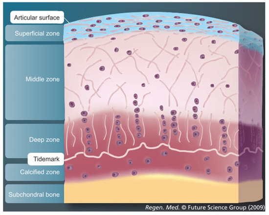
Jfmk | free full-text | ex vivo systems to study chondrogenic ...
Human Eye Diagram: Contrary to popular belief, the eyes are not perfectly spherical; instead, it is made up of two separate segments fused together. Explore: Facts About The Eye To understand more in detail about our eye and how our eye functions, we need to look into the structure of the human eye.

Ch 13 lab map, ch 12 lab map, ch 11 lab map, ch 10 lab map, ch 13 ...
Drag the labels onto the diagram to identify the cells and fibers of connective tissue proper using diagrammatic and histological views. This problem has been ...
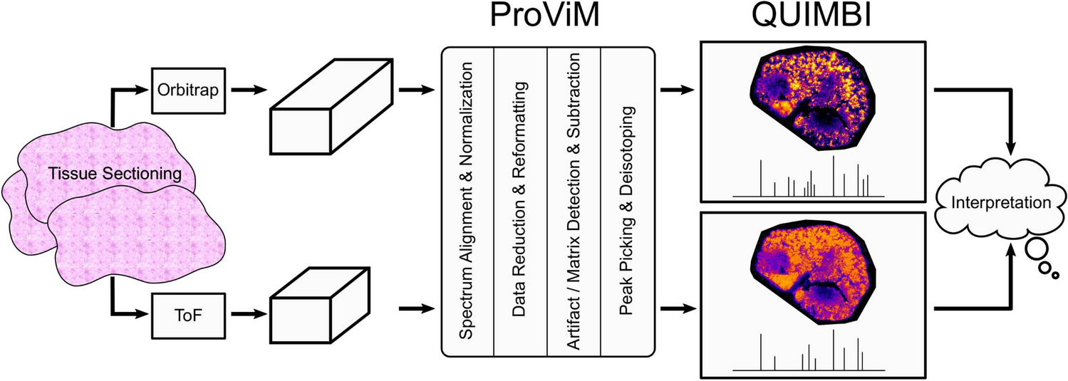
Fast visual exploration of mass spectrometry images with ...
The optical microscope often referred to as the light microscope, is a type of microscope that uses visible light and a system of lenses to magnify images of small subjects. There are two basic types of optical microscopes: Simple microscopes. Compound microscopes. The term "compound" in compound microscopes refers to the microscope having ...

Tissue test study guide flashcards | quizlet
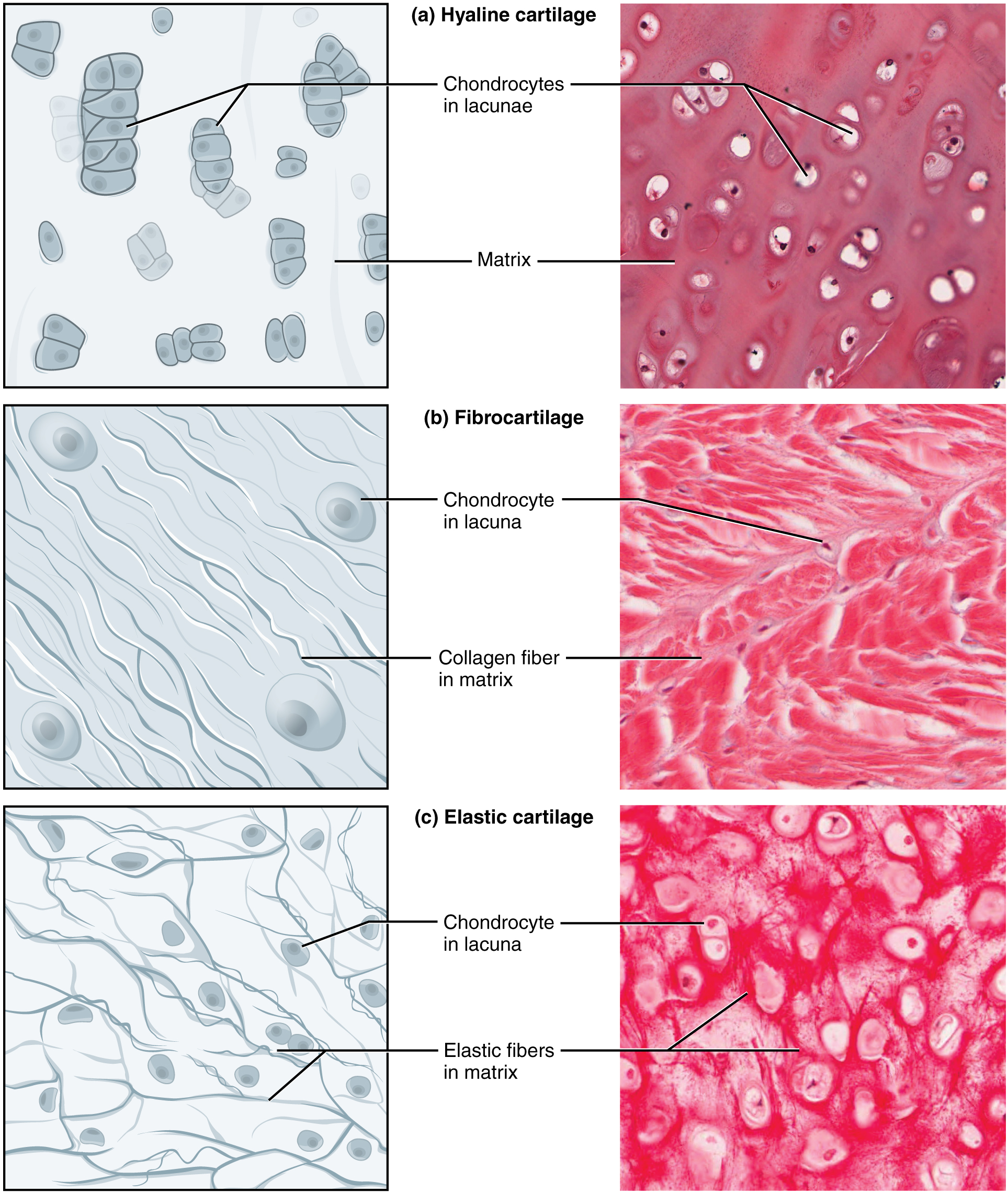
Connective tissue is a tissue that holds the cells together. They provide nourishment, stability, strength, and support to various organs of the body. The six types of connective tissue are bones, cartilage, and loose connective tissue, and adipose tissue, dense regular and irregular connective tissues. Also Read:

Ch 5-7 lab a&p mastering flashcards | quizlet
Depending on the type of cells present (fibroblasts, osteocytes, erythrocytes) and the ECM arrangement, connective tissue can be classified as connective tissue proper or specialized connective tissue. Connective tissue proper is further subdivided into loose connective tissue, mostly found in internal organs as supporting tissue stroma, and ...

Drag the labels onto the diagram to identify the structures and ...
Drag the labels onto the diagram to identify the types of connective tissue proper. look at pic. Image: Drag the labels onto the diagram to identify the ... Rating: 5 · 3 reviews
Connective tissue supports and protects – anatomy and physiology
Hier sollte eine Beschreibung angezeigt werden, diese Seite lässt dies jedoch nicht zu.
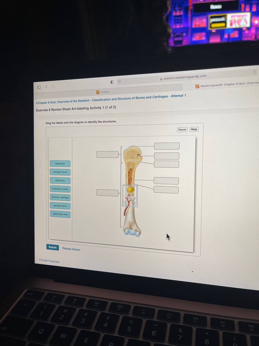
Answered: a session.masteringaandp.com e… | bartleby
Get 24⁄7 customer support help when you place a homework help service order with us. We will guide you on how to place your essay help, proofreading and editing your draft – fixing the grammar, spelling, or formatting of your paper easily and cheaply.
Solved drag the labels onto the diagram to identify the | chegg.com
The study of tissues with a microscope is called ______. histology ... Drag the labels onto the diagram to identify the abdominopelvic regions.

2017 pearson education, inc. - ppt download
Principles and Techiniques of Biochemistry and Molecular Biology 7th ed wilson walker
Solved the cells and fibers of connective tissue proper | chegg.com
Drag the labels onto the diagram to identify the structures and ligaments of the shoulder joint in a newborn the large bones of the skull . This problem has been solved! Shoulder anatomy joint cuff bursa bursitis tendon muscle subacromial arm deltoid diagram ligament acromion blade coracoid humerus inflammation .
Solved drag the labels onto the diagram to identify the | chegg.com
The tissue plays a central role in the experiment and it is important that it is processed so that epitopes and proper morphology is preserved. The most common processing for IHC is to prepare formalin-fixed paraffin-embedded (FFPE) tissue blocks. The purpose of formalin fixation is to produce chemical cross-linking of proteins within the tissue. This terminates all cellular …

Hw 2.pdf - hw 2 due 11:59pm on friday to understand how points are ...
A tissue is a group of cells, in close proximity, organized to perform one or more specific functions. There are four basic tissue types defined by their morphology and function: epithelial tissue, connective tissue, muscle tissue, and nervous tissue. Epithelial tissue creates protective boundaries and is involved in the diffusion of ions and ...

Drag the labels onto the diagram to identify the structures and ...
Four Chambers of the Heart and Blood Circulation. The shape of the human heart is like an upside-down pear, weighing between 7-15 ounces, and is little larger than the size of the fist. It is located between the lungs, in the middle of the chest, behind and slightly to the left of the breast bone. The heart, one of the most significant organs ...
Solved drag the labels onto the diagram to identify the | chegg.com
The pelvis is a group of fused bones and may be considered the first step in the linkage of the axial skeleton (bones of the head, neck, and vertebrae) to the lower appendages. The part of the axial skeleton directly communicating with the pelvis is the lumbar spinal column. The femur is the appendicular skeletal bone connected to the pelvis at the acetabulum, a bony ring formed by the fusion ...
Solved drag the labels onto the diagram to identify the | chegg.com
Drag the labels onto the diagram to identify the types of epithelia.. Signal recognition particle SRP binds to the signal peptide as it emerges from the ribosome. part a drag the labels onto the diagram to identify the part a drag the labels onto the diagram to identify the stages of the life cycle not all labels will be used answer chapter 8 ...
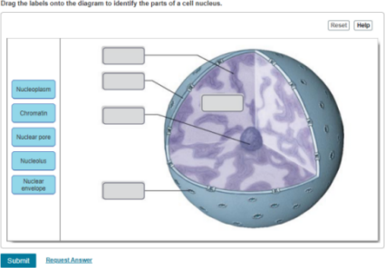
Drag the labels onto the diagram to identify the parts of the cell ...

Ch 5-7 lab a&p mastering flashcards | quizlet

Drag the labels onto the diagram to identify the tissues and ...
A&p chapter 4 tissue: the living fabric flashcards - easy notecards

Nanoparticles influence in skin penetration of drugs: in vitro and ...

Ch 5-7 lab a&p mastering flashcards | quizlet

4.3 connective tissue supports and protects – anatomy & physiology

Drag the appropriate labels to their respective targets. ct ...
Solved -aibrocytes reticular fbers bundles part a drag the | chegg.com

Ch 5-7 lab a&p mastering flashcards | quizlet

Ch 5-7 lab a&p mastering flashcards | quizlet
0 Response to "40 drag the labels onto the diagram to identify the types of connective tissue proper."
Post a Comment