36 sea urchin anatomy diagram
Sea Urchins and Sea Stars. The Sea Urchins and Sea Stars ClipArt gallery includes 139 illustrations of several sea star and sea urchin species. Sea stars, also called starfish, are echinoderms that are in the shape of a star, with typically five arms. Sea urchins are echinoderms that are small, spiny sea creatures shaped like a sphere or globe. Dogfish Internal Anatomy Diagram Wiring Diagram Images Gallery External anatomy of the dogfish shark. The spiny dogfish shark is a small shark that is deep gray with some white spots. A main muscular reservoir that empties after the ventricle contracts. ... Sea Urchin Diagram Cell Model Project Diagram Marine . Pin On Ideas For The House .
sea urchins. In sea urchin. …a complex dental apparatus called Aristotle's lantern, which also may be venomous. The teeth of Aristotle's lantern are typically extruded to scrape algae and other food from rocks, and some urchins can excavate hiding places in coral or rock—even in steel. Sea urchins live on the ocean floor, usually on….

Sea urchin anatomy diagram
Sometimes a sea urchin grows larger than its dugout depression and gets stuck — for life. Once trapped, the urchin can only feed on particles that drift by with the current. Whichever lantern you use to refer to an Urchin's mouth, those teeth are pretty formidable. In areas without a lot of predators (like sea stars and sea otters), purple ... ''Diadema setosum'' is a species of long-spined sea urchin belonging to the family Diadematidae. It is a typical sea urchin, with extremely long, hollow spines that are mildly venomous. ''D. setosum'' differs from other ''Diadema'' with five, characteristic white dots that can be found on its body. The species can be found throughout the Indo-Pacific region, from Australia and Africa to Japan ... Sea Urchin Dissections 1. Wear new latex gloves at all times a. The human hands contain RNA degrading enzymes that can digest delicate RNA molecules 2. Pre-label the cryovials with a. Date of dissection b. Urchin genus and species name c. Collection location d. Tissue type (example: digestive, intestine, stomach, etc) e. ID number (example ...
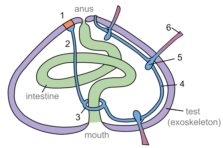
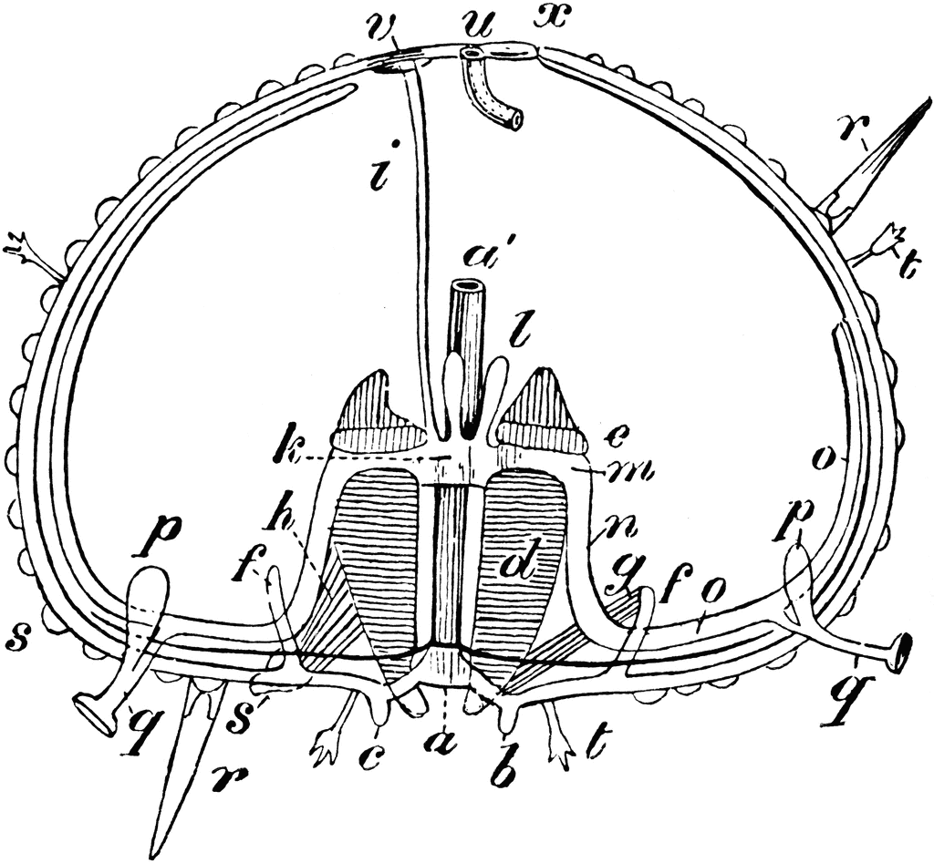
Sea urchin anatomy diagram. The video will teach you how to dissect a green & purple sea urchin step by step, and review the external and internal anatomy of the sea urchin. In this video, we'll cover the following structures: Epidermis Aristotle's lantern. Tube feet Pharynx. Spines Esophagus. Tubercles Stomach. Mouth Intestine. Teeth Mesentery tissue. Sea Urchin Anatomy study guide by Thomas_Schroeder6 includes 17 questions covering vocabulary, terms and more. Quizlet flashcards, activities and games help you improve your grades. Internal Anatomy of the sea urchin The inside of a sea urchin is dominated by a large, coiled digestive system which consists basically of a tube joining the lower mouth to the anus on the upper surface. In regular echinoids the mouth opens into an oesophagus (oe) that initially runs through the centre of the Aristotle's lantern. A thorough understanding of the sea urchin (Echinodermata: Echinoidea) digestive tract anatomy is a prerequisite for the correct interpretation of physiological and biomechanical analyses focusing ...
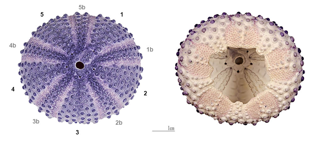
Download scientific diagram | External anatomy of a sea urchin based on a specimen of Strongylocentrotus purpuratus. A shows the numbering of the genital plates. The plates indicated by dotted ... Sea urchins (/ ˈ ɜːr tʃ ɪ n z /) are spiny, globular echinoderms in the class Echinoidea.About 950 species of sea urchin live on the seabed of every ocean and inhabit every depth zone — from the intertidal seashore down to 5,000 metres (16,000 ft; 2,700 fathoms). The spherical, hard shells of sea urchins are round and spiny, ranging in diameter from 3 to 10 cm (1 to 4 in). Sea Urchin Anatomy One look at a sea urchin and you can see why they would be called sea hedgehogs. They have hard rounded shells covered with sharp movable spines. Urchins are part of the phylum Echinoderm and their name comes from Ancient Greek (echinos meaning "hedgehog" and derma meaning "skin"). There are more than 900 species of sea urchins and they come in a range of the body that are not addressed by this muscle anatomy diagram, however the muscles that are of primary interest from a fitness and exercise Aug 22, 2015 · Sea Urchin Anatomy One look at a sea urchin and you can see why they would be called sea hedgehogs. They have hard rounded shells covered with sharp movable spines. Urchins are part
Sea urchins have no "brain" as we think of it, instead they have a nerve ring near the hydraulic system used to power the tube feet. THE TUBE FEET. The tube feet are the primary means of locomotion for the sea urchin (along with moving the spines). Lateral movement is accomplished by means of paired muscles in the tube foot itself and extension and retraction by muscles in the hydraulic "bulb" behind each tube foot. While each "foot" may seems weak and ineffective, the large numbers of feet ... 1 Ectoderm, 2 Archenteron (central), 3 Endoderm, 4 First group of mesoderm cells, 5 Origine of the first group of migrating mesoderm cells, 6 Origine of the second group of mesoderm cells, 7 Invagination area. Pluteus larval stage. Information: The final gastrula gives rises to the pluteus larva in the sea urchin. Diagram the anatomy with this printable. Purple Sea Urchin Printable - Purple sea urchin printable diagram anatomy sheet. Sand Dollar Printable - Sand dollars are beautiful sea animals. Learn about what they eat and their predators with this informational sheet. Sea Cucumbers Printable - Printable diagram and labeling sheet of a sea cucumber. Small sea urchins species have spines a few cms long at most. The long-spine variety exceed 10 cms, and the largest may have spines up to 30 cms (= 1 foot) long. The length, as shown here, means that when the creatures are safely lodged in crevices near their algae supply, their spines remain very visible. Anatomy of a long-spined sea urchin.
Basic Tissue Layers for Planarian Cross Section. Tapeworm Anatomy. Life Cycle of a Beef Tapeworm. Rotifer Parthenogenesis Diagram. Anatomy of a Nemertea (not used in summer) Circulatory System of a Nemertea. Anatomy of a Chiton. Phylogeny of Spiralia Phyla for ZO 150. Circulatory System of a Polychaeta Metamere.
(Click to Expand): Diagram of the basic anatomy of a "regular" sea urchin. Click to Expand: Cucumariid Sea Cucumber (Simple organ diagram) Starfish (Echinodermata Phylum) Asterias Star Fish (Echinodermata Phylum) Starfish and sea stars like this are known as echinoderms that are from the class Asteroidea. Starfish similar to other echinoderms ...
The average sea urchin is 3 to 10 centimeters in diameter. The biggest known sea urchin species is 36 centimeters in diameter. Physical Characteristics Sea urchins do not have a recognizable face, but they have an identifiable mouth and anus. They exhibit radial symmetry: they have five sections in equal size.
Chapter 14. Gastrulation and Neurulation. "It is not birth, marriage, or death, but gastrulation , which is truly the most important time in your life." During gastrulation, cell movements result in a massive reorganization of the embryo from a simple spherical ball of cells, the blastula, into a multi-layered organism.
In this article we will discuss about the structure of Sea Urchin (Echinus) with the help of a diagram. 1. It is commonly known as “sea urchin” and is formed in shallow water in both rocky and sandy place in sea. 2. Body is Sub-globular and convex or dome-shaped above and flattened below. The aboral and oral surfaces are distinct.
How the Purple Sea Urchin Digests Food. 1. The food enters the mouth, which unlike most animals, is located at the bottom of the organism. 2. The food is then mechanically digested by Aristotle's Lantern which is an arrangement of teeth, muscles, and plates. 3.
Purple sea urchin is spiny and ovoid shaped. Its upper surface is domed and the underside is flattened. The mouth has a complex structure made up of five calcium carbonate plates. It is referred to as "Aristotle's lantern" because he was the first to describe it. This sea urchin grows from a greenish colored juvenile to a vivid purple adult.
The sea urchin reproductive system is an organ system responsible for the production of gametes that will eventually be released into the sea for spawning. Almost all sea urchins have separate sexes and release either eggs or sperm into the sea in the spring, which is their breeding time.
INTERNAL ANATOMY. From The Fishery Biology of Sea Urchins in the Family Strongylocentrotidae, Washington Department of Fisheries, 1976. If you cut the sea urchin along the equator you will see the following: Am and Im are the ambulacral and interambulacral areas respectively. A is the "top" and B is the bottom or mouth side.
How Sea Urchins Move This purple urchin moves along the bottom of the aquarium in much the same way it would in the ocean. You can see some of the tube feet attached to the rocks as it pulls itself along. The tube feet are part of the urchin's water vascular system. Urchins can also move using their spines, which are controlled by strong muscles.
urchin anatomy. about us. teacher resources. useful links. Select Language: Our Acidifying Ocean. Part 1 Part 2 Part 3 Part 4. Learn Experiment Analyze Act. Close. Problems with this site? Click here. ©2008-2020, University of Washington. Sponsored by the National Science Foundation.
Sea Urchin Dissections 1. Wear new latex gloves at all times a. The human hands contain RNA degrading enzymes that can digest delicate RNA molecules 2. Pre-label the cryovials with a. Date of dissection b. Urchin genus and species name c. Collection location d. Tissue type (example: digestive, intestine, stomach, etc) e. ID number (example ...
''Diadema setosum'' is a species of long-spined sea urchin belonging to the family Diadematidae. It is a typical sea urchin, with extremely long, hollow spines that are mildly venomous. ''D. setosum'' differs from other ''Diadema'' with five, characteristic white dots that can be found on its body. The species can be found throughout the Indo-Pacific region, from Australia and Africa to Japan ...
Sometimes a sea urchin grows larger than its dugout depression and gets stuck — for life. Once trapped, the urchin can only feed on particles that drift by with the current. Whichever lantern you use to refer to an Urchin's mouth, those teeth are pretty formidable. In areas without a lot of predators (like sea stars and sea otters), purple ...
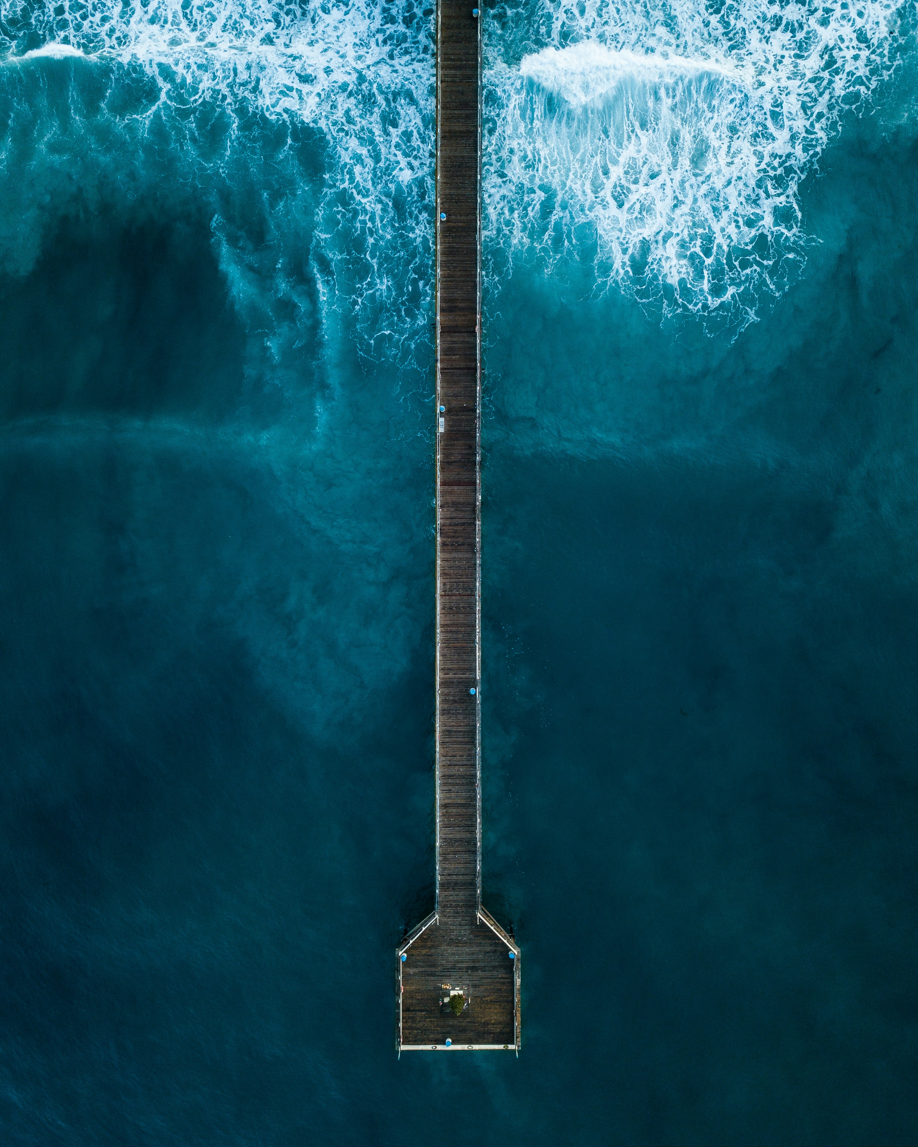
Early in the morning along the Southern California coast in San Diego, we were exploring the vast and beautiful beach neighborhood of Pacific Beach. There were a ton of surfers early in the morning as the sun was rising, but the pier was completely empty. This was an amazing and rare change to capture something at a time that it wasn’t full of people and the hustle and bustle of life. It was extremely calming just listening to the ocean waves and watching the sunrise.
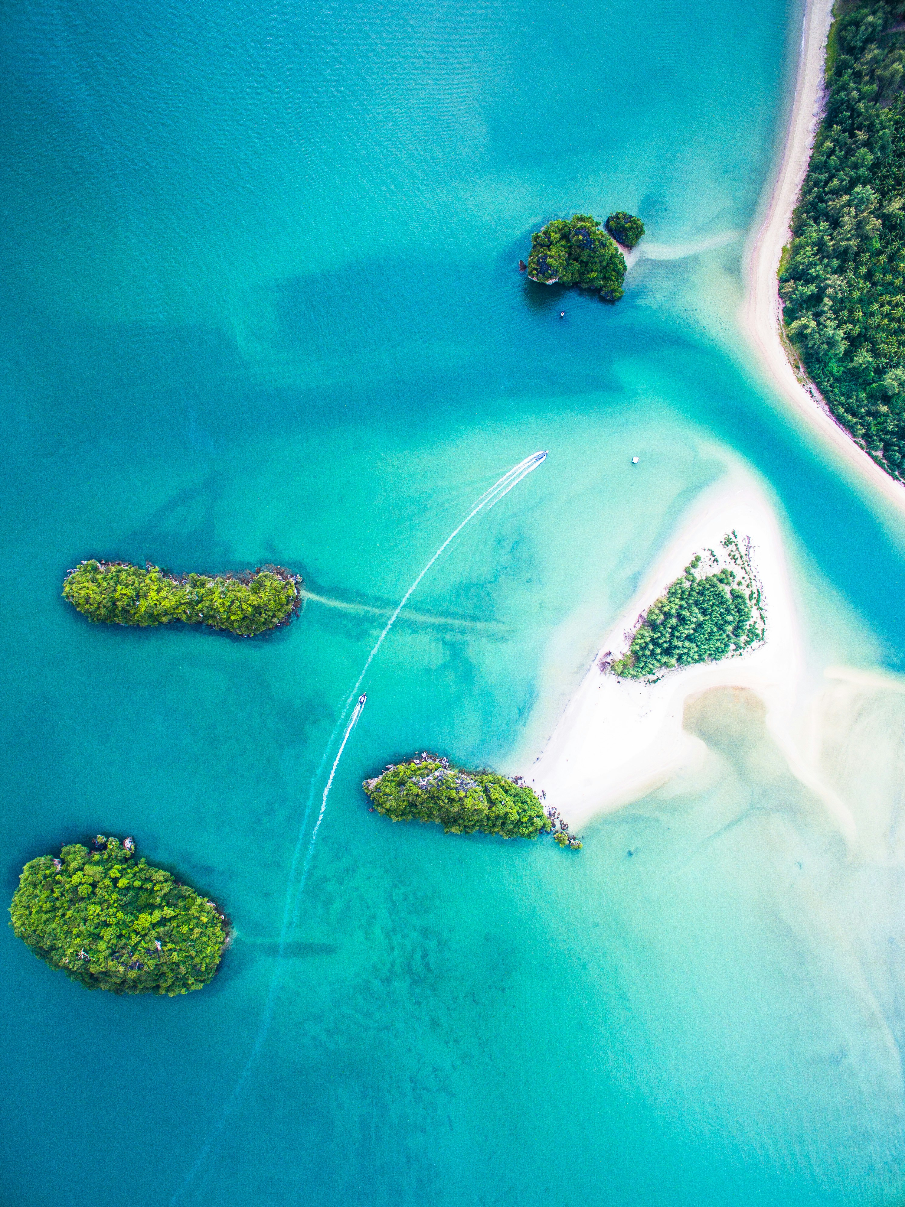
This photo was shot with my DJI Phantom 3 in Thailand, Krabi in 2016. It’s possible to get to the Sirithan Beach (sandy island) walking from the mainland (more right) in the water. The maximum depth in the most shallow path is waist-high. A little inconvenience totally worth it because the beach is almost empty and feels like a paradise.
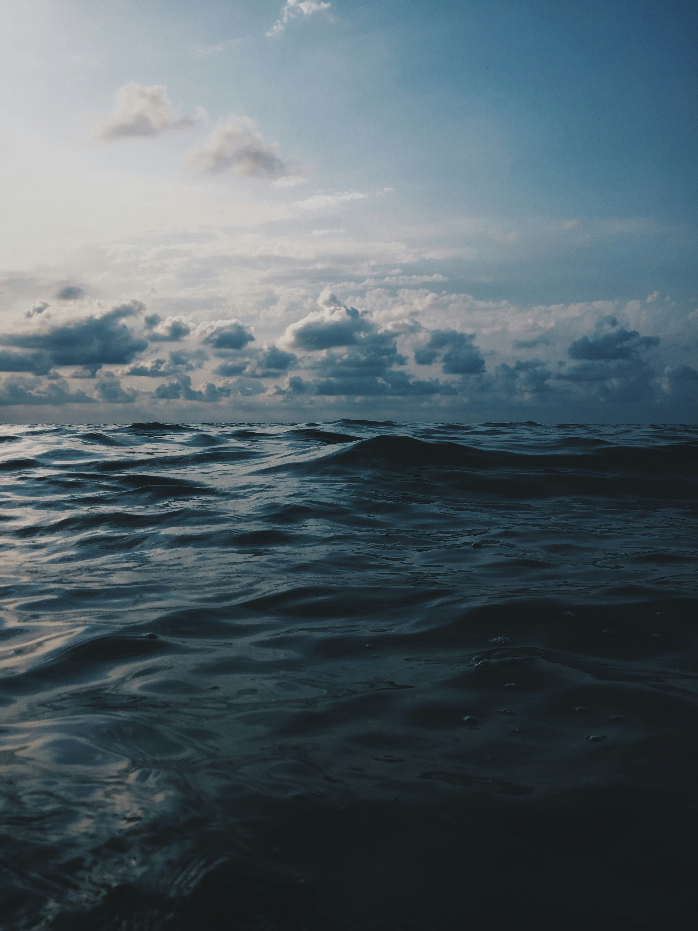
To cut a long story short I was swimming and suddenly decided to take a photo. why? because when I was swimming I could see the beauty of the black sea with nice clouds and though I’ve to save this moment. So, I took my iPhone from the beach, went into the sea, open the camera and took the photo and here it is what it turned out.






0 Response to "36 sea urchin anatomy diagram"
Post a Comment