37 acl mcl pcl lcl diagram
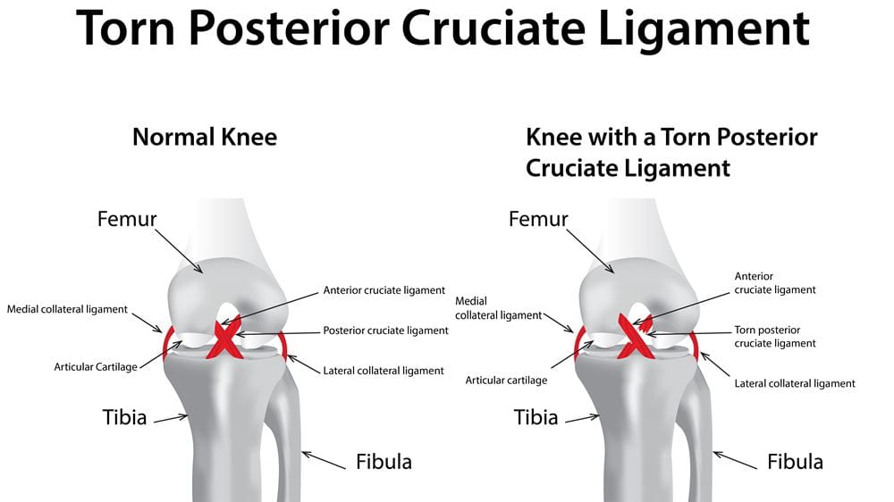
Injury: Abnormal passive posterior displacement, posterior drawer sign. Anterior Cruciate Ligament (ACL) Attachments: from anterior tibia to lateral femoral condyle anteriorly. Alignment: perpendicular to lateral collateral ligament and PCL (slightly shorter than PCL) Function: Key stabilizer of knee joint. Check posterior movement of femur on ... Posterior cruciate ligament (PCL) injury happens far less often than does injury to the knee's more vulnerable counterpart, the anterior cruciate ligament (ACL). The posterior cruciate ligament and ACL connect your thighbone (femur) to your shinbone (tibia). If either ligament is torn, it might cause pain, swelling and a feeling of instability.
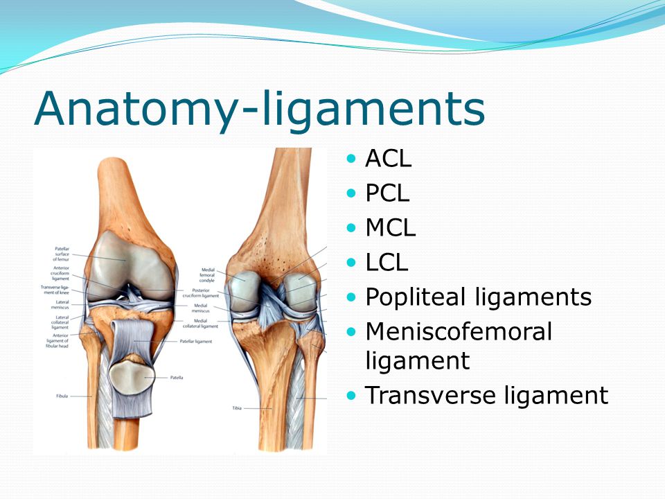
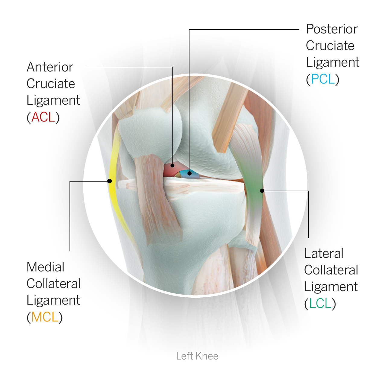
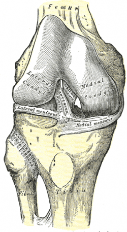
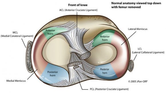
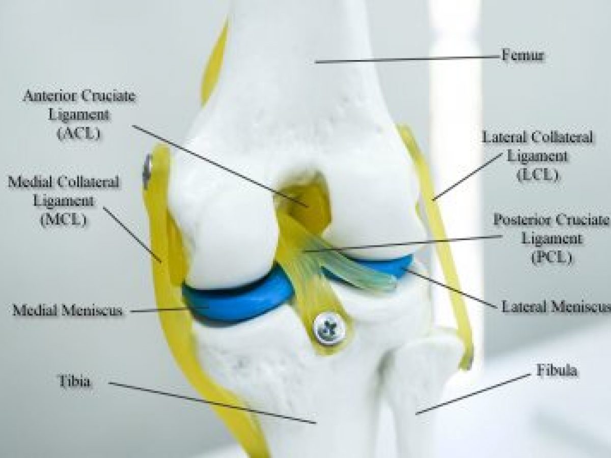
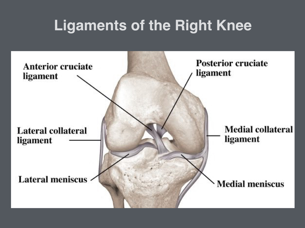
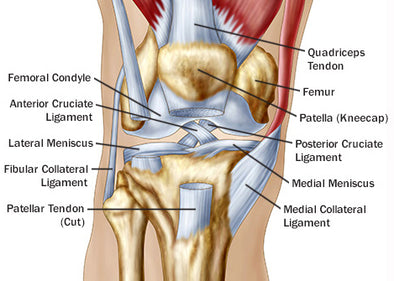
In the model you can see femur, tibia and fibula, with ACL (anterior cruciate ligament), PCL (posterior cruciate ligament), MCL (medial collateral ligament), LCL (lateral collateral ligament) and medial and lateral meniscus. These are the most typical ligament and cartilage structures in the knee to receive an injury.

Acl mcl pcl lcl diagram
anterior cruciate ligament acl injuries symptoms, what is the mcl the knee, acl solutions acl knee anatomy and diagram images, anatomy of the knee « acl recovery secrets, acl injury symptoms and causes mayo clinic, acl mcl pcl meniscus pinterest, instrumented arthrometry for diagnosing partial versus, basic science of anterior cruciate ... Acl And Mcl Diagram. Signs and symptoms of a medial collateral ligament (MCL) injury include The anterior cruciate ligament (ACL) and posterior cruciate ligament. The MCL is one of four major ligaments that supports the knee. either partial or complete ruptures in the ligament significantly increases the load on the ACL. Posterior Cruciate Ligament (PCL) Posterior Drawer Test. This test is performed with the patient supine and the knee flexed to 90°. There are two different ways it may be performed. The first is the opposite of the anterior drawer test. Absence of a discrete endpoint with posterior force applied to the tibia is considered positive.
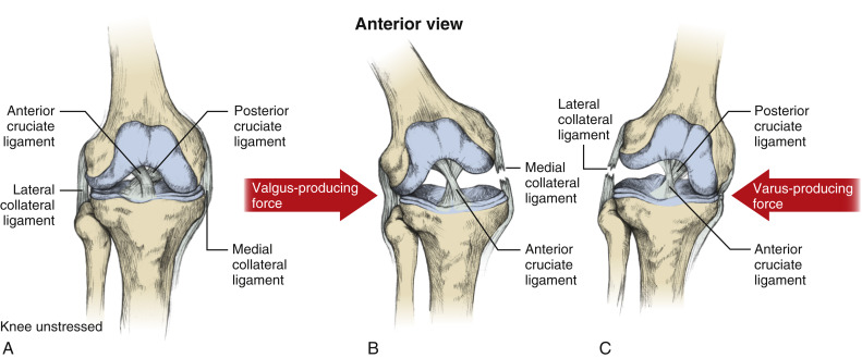
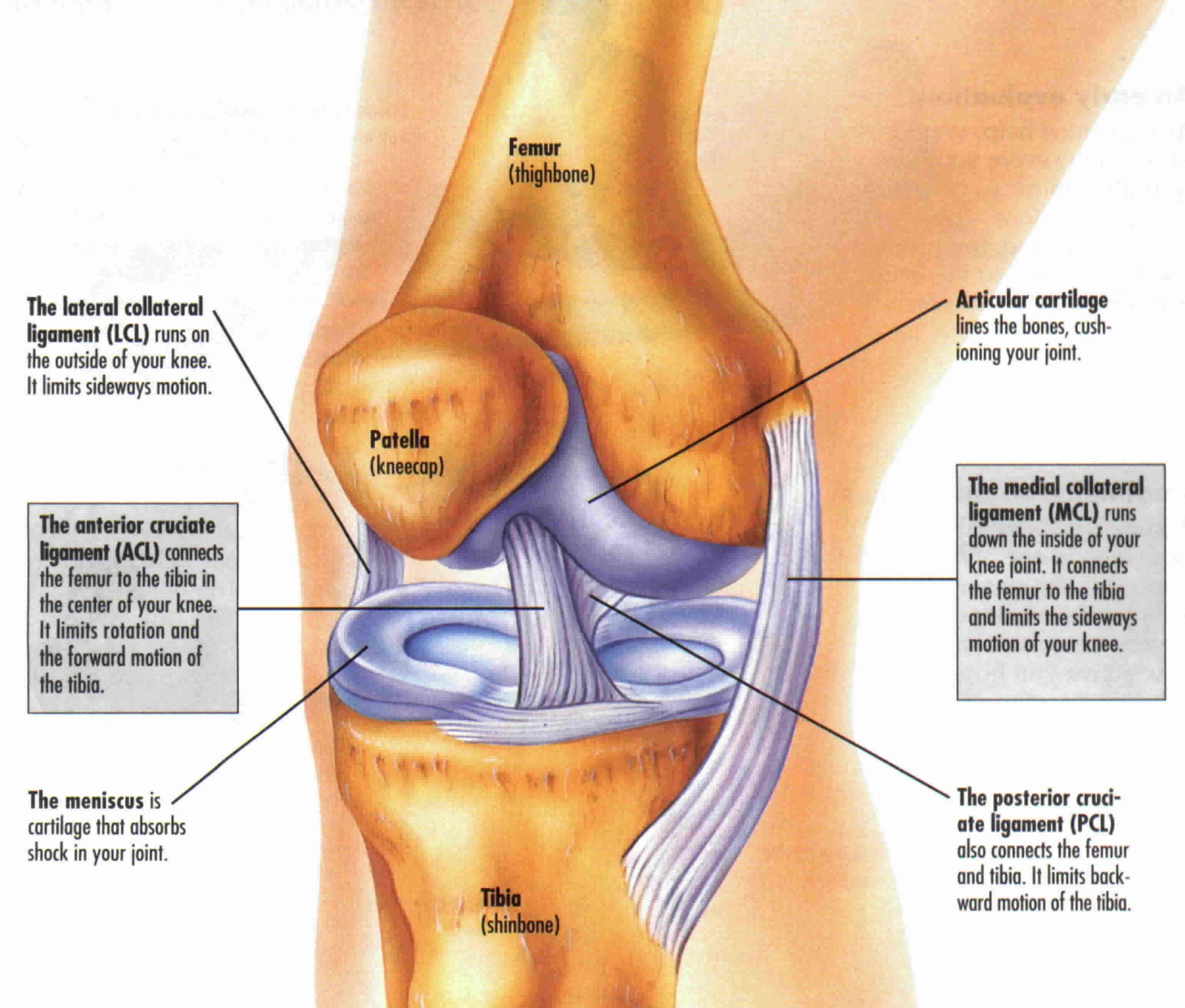
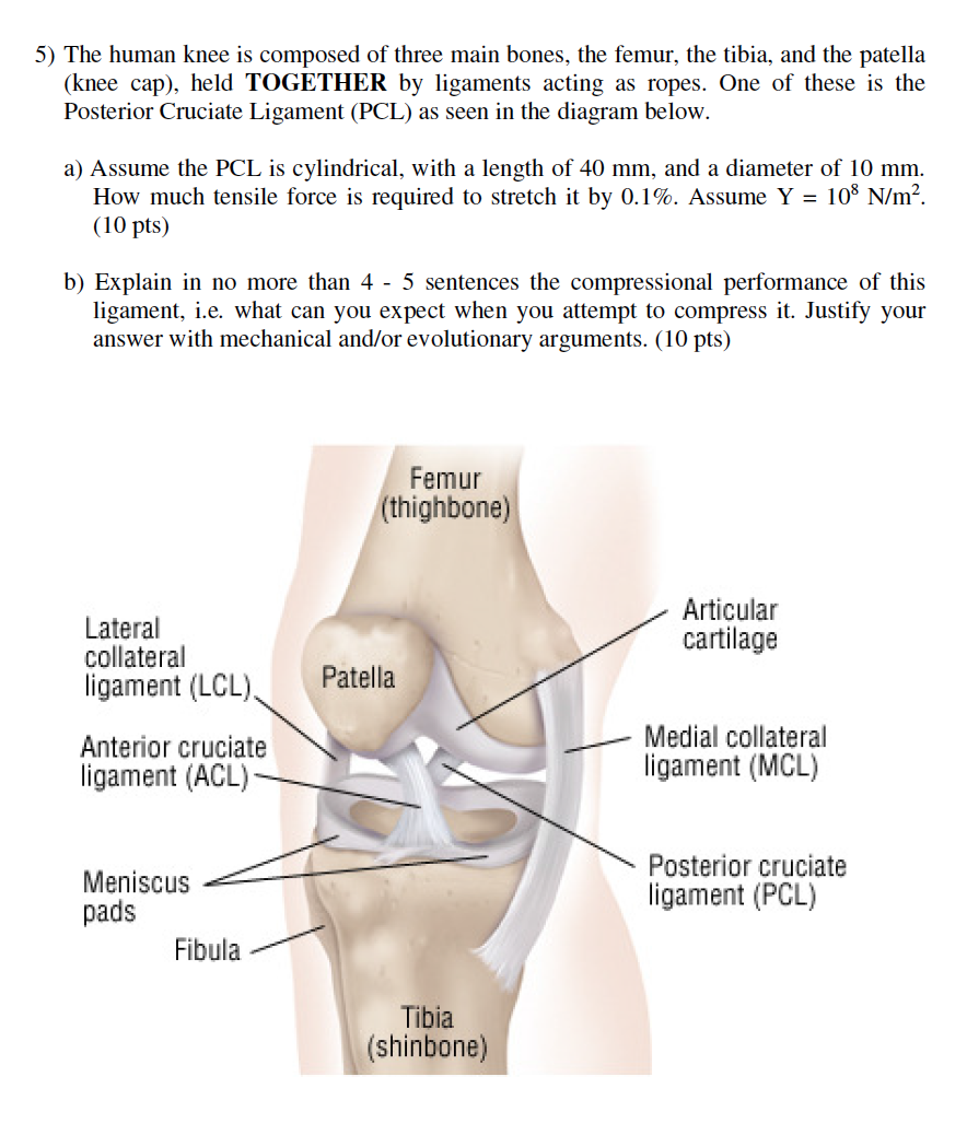
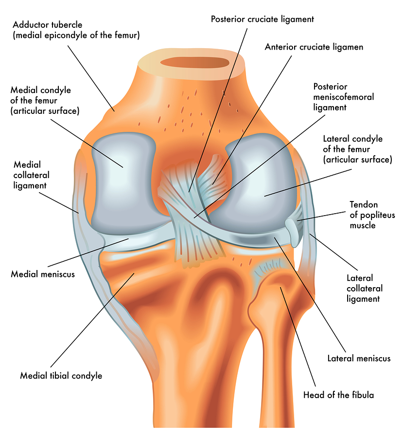
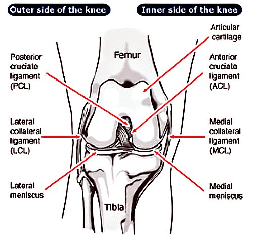
Acl mcl pcl lcl diagram. The ACL prevents the tibia from sliding forward along the femur, while the PCL prevents the tibia and femur from sliding towards each other. The other two ligaments of the knee, the medial collateral ligament (MCL) and lateral collateral ligament (LCL). These run along the outside of the knee and prevent the knee from bending sideways. Knee ligaments are bands of tissue that connect the thigh bone in the upper leg to the lower leg bones. There are four major ligaments in the knee: ACL, PCL, MCL and LCL. Injuries to the knee ligaments are common, especially in athletes. A sprained knee can range from mild to severe. Talk to a healthcare provider if you have a severe knee ... staged for optimal outcomes. Surgery uses an allograft or autograft to reconstruct the torn ACL and PCL ligaments, and may repair the MCL, LCL, and/or posterolateral corner of the knee if needed as well. Long-term complications after surgery include chronic pain, knee instability, arthrofibrosis, and loss of knee flexion ROM. the Posterior Cruciate Ligament (PCL) and Anterior Cruciate Ligament (ACL) control back-and-forth movement; the Medial Collateral Ligament (MLC) helps to brace the inside of the knee; the Lateral Collateral Ligament (LCL) braces the ouside of the knee, controlling sideways motion and protecting the knee from over-extending. While most injuries ...
the femur thigh bone in the upper leg and the tibia shinbone in the, knee lints acl mcl diagram 11 18 tierarztpraxis ruffy de u2022 rh parison of strain in knee lints acl pcl mcl and lcl diagram of a knee inspirational acl pcl mcl acl vs mcl pcl absolute life wellness center what is an acl pcl and functions The diagram illustrates where the fours main knee ligaments are located; the anterior cruciate ligament (ACL), the posterior cruciate ligament (PCL), the medial collateral ligament (MCL), and the ... Medial Collateral Ligament - MCL. prevents inward or medial collapse. Meniscus. provides stability and cushion. ... Skeleton Diagram. 8 terms. Bitter_Breezy. OTHER SETS BY THIS CREATOR. Fractures. 14 terms. ... Posterior Cruciate Ligament -PCL. prevents the tibia from shifting posterior. Anterior Cruciate Ligament - ACL ... Ligaments Of The Knee. The four main ligaments in the knee are: Medial Collateral Ligament: found on the inner side of the knee Lateral Collateral Ligament: found on the outer side of the knee Anterior Cruciate Ligament: found in the middle of the knee Posterior Cruciate Ligament: found in the middle of the knee The collateral and cruciate ligaments are generally considered to be the main ...
The anterior cruciate ligament (ACL) is one of a pair of cruciate ligaments (the other being the posterior cruciate ligament) in the human knee.The two ligaments are also called "cruciform" ligaments, as they are arranged in a crossed formation. In the quadruped stifle joint (analogous to the knee), based on its anatomical position, it is also referred to as the cranial cruciate ligament. Ligament Injury (ACL, PCL, MCL, LCL & multiligament injury) ... Diagram of Anterior Cruciate Ligament - Knee Surgeon Melbourne ... Download scientific diagram | Concomitant ligament injuries (ACL anterior cruciate liament , MCL medial cruciate liament, PCL posterior cruciate lia- ment) from publication: Lateral ligament ... Anterior cruciate ligament (ACL) Posterior cruciate ligament (PCL) Medial Collateral ligament (MCL) Lateral collateral ligament/Posterolateral Corner (LCL/PLC) This specific rehabilitation protocol should be used when both the ACL and PCL are reconstructed, along with one or more of the other ligaments.
The lateral collateral ligament runs along the outside of your knee and prevents it from moving too far out. Without the MCL or LCL your knee will feel wobbly, especially if you do a side-to-side cutting motion. Thankfully, neither the MCL nor the LCL are removed during a total knee replacement surgery. However, on rare occasions, the MCL can ...
Title: Diagram Of Acl Author: OpenSource Subject: Diagram Of Acl Keywords: diagram of acl, common knee injuries orthoinfo aaos, configuring ip access lists cisco, acl rehabilitation when is your athlete really ready for, what is an acl pcl and their functions sports knee, anterior cruciate ligament acl rehabilitation physiopedia, clinical diagnosis of an anterior cruciate ligament, knee ...
Anterior cruciate ligament (ACL) is the most commonly injured knee ligament.It connects the thigh bone to the shin bone. Posterior cruciate ligament (PCL) also links the thigh bone to the shin ...
30.9.2021 · N. Korea's parliamentary session. This photo, released by North Korea's official Korean Central News Agency on Sept. 30, 2021, shows Kim Yo-jong, North Korean leader Kim Jong-un's sister and currently vice department director of the ruling Workers' Party's Central Committee, who was elected as a member of the State Affairs Commission, the country's …
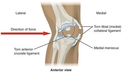
The medial collateral ligament (MCL) is a flat band of connective tissue that runs from the medial epicondyle of the femur to the medial condyle of the tibia and is one of four major ligaments that supports the knee. MCL injuries often occur in sports, being the most common ligamentous injury of the knee, and 60% of skiing knee injuries involve the MCL).
29 Dec 2020 — These are the four main ligaments or tough bands of tissue in the knee. ACL is the anterior cruciate ligament, which connects the thigh bone to ...
ACL vs. MCL tears: Although symptoms of ACL and MCL tears are similar, a few key differences will help identify whether the injury affected the ACL or MCL. An ACL tear will have a more distinctive and loud popping sound than an MCL tear. The location of your pain and swelling could indicate either an ACL or MCL tear.
MCL Tear Symptoms. Just as with an ACL tear, you may hear a popping sound when the injury occurs. Some of the most common symptoms of an MCL tear include: Pain on the inner side of the knee. Feeling the knee "give out". Swelling. The knee feels unstable. Pain when putting weight on the knee. Locked knee.
The posterior cruciate ligament (PCL) runs diagonally opposite of the ACL, crossing its path to make an “X” in the middle of the knee. Unlike the ACL, the PCL restricts excessive posterior translation, or sliding backward of the tibia on the femur. This ligament is nearly twice as thick as the ACL, meaning that it is almost twice as strong.
A knee ligament injury a sprain of one or more of the four ligaments in the knee, either the Medial Collateral Ligament (MCL), Lateral Collateral Ligament (LCL), Posterior Cruciate Ligament (PCL), or the Anterior Cruciate Ligament (ACL). ACL, PCL, MCL, and LCL injuries are caused by overstretching or tearing of a ligament by twisting or wrenching the knee.
Different Types Of Knee Injuries – ACL, LCL, MCL and PCL. Our knees are one of the most vital joints in our bodies, shouldering our weight and facilitating movements like walking, running and sitting. Your knee is made up of four distinct ligaments, with one on the front, back and each side. Damage to the knee can result in a partial or ...
Abbreviations: Medial collateral ligament (MCL) Anterior cruciate ligament (ACL) Posterior cruciate ligament (PCL) Lateral collateral ligament (LCL) Injury ACL N 151 39% ACL + MCL 118 30% ACL + posterior 1 0% medial capsule MCL 47 12% Posterior medial 12 3% capsule ACL + LCL LCL 7 2% 2 1% + PCL 19 5% MCL + PCL 8 2%
Posterior Cruciate Ligament (PCL) Posterior Drawer Test. This test is performed with the patient supine and the knee flexed to 90°. There are two different ways it may be performed. The first is the opposite of the anterior drawer test. Absence of a discrete endpoint with posterior force applied to the tibia is considered positive.
Acl And Mcl Diagram. Signs and symptoms of a medial collateral ligament (MCL) injury include The anterior cruciate ligament (ACL) and posterior cruciate ligament. The MCL is one of four major ligaments that supports the knee. either partial or complete ruptures in the ligament significantly increases the load on the ACL.
anterior cruciate ligament acl injuries symptoms, what is the mcl the knee, acl solutions acl knee anatomy and diagram images, anatomy of the knee « acl recovery secrets, acl injury symptoms and causes mayo clinic, acl mcl pcl meniscus pinterest, instrumented arthrometry for diagnosing partial versus, basic science of anterior cruciate ...














/knee-pain-instability-2549493-5c04aaf946e0fb00010b8e7a-b0ef89c536aa4cd7a9db3f927d72597b.png)



0 Response to "37 acl mcl pcl lcl diagram"
Post a Comment