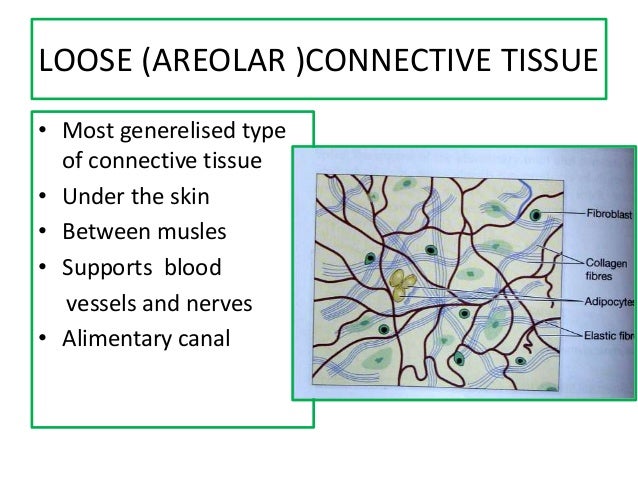
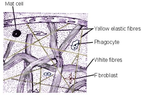
38 areolar connective tissue diagram
I need to find how these cells have adapted to suit their functions(in detail), I have never done biology before and this is my homework for Forensics, literally my worst science, help me out biologists. A magnified image of areolar connective tissue highlights its mesh-like appearance, with the protein fibers visible as a ...
This tissue is present in the skin. Cartilage, bones, and blood are various types of specialized connective tissues. 4. Adipose and blood tissue Adipose tissue: Adipose tissue is another type of loose connective tissue located mainly beneath the skin. The cells of this tissue are specialized to store fats.
Areolar connective tissue diagram
ANS = Areolar connective tissue is made of cells and extracellular matrix ("extra-" means "outside", so the extracellular matrix is material ... There are 4 basic types of fabrics: Connective tissue, epithelial tissue, muscle tissue, and nervous tissue.Connective tissue supports and connects other tissues (bone, blood, and lymphatic tissue). Epithelial tissue forms a covering (skin, lining of various body ducts). This layer is protective of the submucosa and mucosa, as well as helps to move food through the stomach. 4. Serosa. This outermost layer of the stomach is a thin membrane that protects the stomach from other organs and the motion of the food inside. It is a thin membrane made up of areolar connective tissue and squamous epithelial tissue.
Areolar connective tissue diagram. There are 4 basic types of fabrics: Connective tissue, epithelial tissue, muscle tissue, and nervous tissue.Connective tissue supports and connects other tissues (bone, blood, and lymphatic tissue). Epithelial tissue forms a covering (skin, lining of various body ducts). Just finished a 10 week RAD cycle and during the cycle I noticed my muscles were growing and repairing faster than my ligaments and tendons and from that my joints (mainly shoulders and elbow joints) weren’t able to keep up with the increase in weight I was able to lift. What remedies do you guys use to maintain healthy and fully functioning joints? (I eat heaps as rad has made me a lot hungrier, drink plenty of water, take my vitamins and stretch plenty) Areolar connective tissue is found between the skin and muscles, around blood vessels and nerves and in the bone marrow. It fills the space inside the organs, supports internal organs and helps in repair of tissues. This type of tissue contains many cells, a loose arrangement of fibres, and moderately viscous fluid matrix. diagram of loose connective tissue. Dense Irregular ...
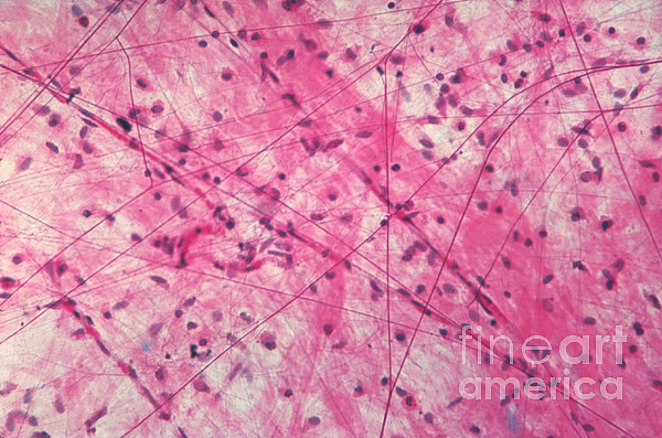
Photomicrograph: Areolar connective tissue, a soft packaging tissue of the body (300x). Epithelium Lamina propria Fibroblast nuclei Elastic fibers Collagen fibers The Skin | Boundless Anatomy and Physiology CHAPTER 1 Introduction The skin is the largest organ of the body, accounting for about 15% of the total adult body weight. Can anyone share experiences about using MM to heal complex autoimmune conditions, such as connective tissue disease? (Specifically, lupus, RA, polymyositis, scleroderma, MCTD). I see a lot of healing about skin conditions and gut issues, but I see far fewer testimonials about healing of very complex conditions. Thanks in advance for sharing. I have suspected for a bit that I am hypermobile - I didn't realize until this year that hypermobility was actually a thing, until I wanted to do flexibility training for circus arts, and learned from a PT I follow online that hypermobile aerialists cannot stretch the same way that "normal" people do. Today I went into the rheumatologist's office and learned that I am actually a full 9 on the beighton scale. I had researched the criteria for hEDS and also read from some studies that hypermobil... Areolar connective tissue. D. Hyaline cartilage. E. Cardiac muscle tissue. 12. From the left ventricle, where does blood pass? A. Right atrium. B. Right ventricle. C. Bicuspid valve. D. Aortic semilunar valve. E. Pulmonary trunk. 13. This structure temporarily shunts blood from the pulmonary trunk into the aorta in a fetus. A.
31.07.2019 ... Type. Location. Characteristics. Function. Areolar tissue. Found beneath the epithelia. Found between skin and muscles, around blood vessels ... Occipital Bone Anatomy Function And Treatment. Occipital Bone Anatomy Function And Treatment. By dubaikhalifas On Jan 19, 2022 A tissue is a: (a) Group of separate organs that are coordinated in their activities. (b) Group of similar cells that function together in a specialized activity. (c) Layer of cells surrounding an organ. (d) Sheet of cells, one layer thick. (b) Group of similar cells that function together in a specialized activity. (a) Neuron: Areolar tissue, blood and tendon are connective tissues while neuron is a part a nervous tissue. (b) Cartilage: RBC, WBC and platelets are parts of vascular connective tissue while cartilage is skeletal connective tissue. (c) Ligament: Ligament is a connective tissue.
Your body is held together by tissues that connect all the structures in your body. When you have connective tissue disease these surrounding structures are effected ( penis). Connective tissue is made up of collegen and elastic, when they are inflamed they can make muscles and organs of the body very stiff. They can get inflamed from injury or repetitive use. There are over 200 connective tissue diseases. As the area gets inflamed the connective tissue is damaged and can eventually cause fibros...
Why were people like Benji,Tessa,Sarah,Annalise,Pia,Janine,Kristie not cast ?
Mods: Please let me know if it's wrong to post this here and you know somewhere else that might help! I'll preface: I don't have a diagnosis of MCTD. I have suspect connective tissue symptoms, and I'll be seeing a rheumatologist in early January. I recently had a surgery where a cyst was removed. It ended up being a super rare kind of cyst, but through reading all the scientific journal articles present (literally starting in 1920-- there are very few). Most articles state that because of the ...
It has four layers. From deep to superficial they are: mucosa (pseudostratified ciliated columnar epithelium and an underlying lamina propria), submucosa (areolar connective tissue with seromucous glands and ducts), hyaline cartilage (16-20 incomplete rings stacked horizontally), and adventitia (areolar connective tissue). 4.
By Benjamin Noé 5 janvier 2022. There are 4 basic types of fabrics: Connective tissue, epithelial tissue, muscle tissue, and nervous tissue. Connective tissue supports and connects other tissues (bone, blood, and lymphatic tissue). Epithelial tissue forms a covering (skin, lining of various body ducts). Contenu cacher.
I’m wondering how long it takes to determine which auto immune disease it is or if it even matters. Also wondering how long you all were on methotrexate. I read about all of these symptoms posted here and it breaks my heart for her. I wish you all the very best.
I’ve unfortunately had multiple injuries in the past six months that have led to knee, ankle and hip tendinitis. I also recently found out that I have some mild bulging in the discs in my lower neck. I’ve never had any sprains or injuries like this, and I was very active as a child doing dance, gymnastics and track. I was never naturally flexible. I saw a rheumatologist who suggested a connected tissue issue, but the only joints in my body that seem to be hyper mobile are my ankles and elbows. ...
About 5 years ago I started having widespread pain. I also had psoriasis and was given a psoriatic arthritis diagnosis eventually. I’ve never had evidence of joint damage on imaging other than “wear and tear arthritis, stenosis, mild to moderate disk bulging”. Many of my symptoms fit enthesitis from PsA; swelling and pain around the joints in my spine, hips, hands, etc. But one of the symptoms that cause me the most pain has never been acknowledged or explained, and that is these ropy lumps ...
By Benjamín Noah Enero. There are 4 basic types of fabrics: Connective tissue, epithelial tissue, muscle tissue, and nervous tissue. Connective tissue supports and connects other tissues (bone, blood, and lymphatic tissue). Epithelial tissue forms a covering (skin, lining of various body ducts). Contenido ocultar.
Here are a number of highest rated Reticular Connective Tissue Drawing pictures upon internet. We identified it from obedient source. Its submitted by admin in the best field. We take on this nice of Reticular Connective Tissue Drawing graphic could possibly be the most trending subject in the same way as we allowance it in google gain or facebook.
The branches course infero-medially within the subcutaneous tissue to effect anastomoses with branches of the internal mammary and intercostal arteries in the areolar area. Because there is often more subcutaneous tissue laterally than medially, they are frequently found from 1 to 2.5 cm from the skin surface.
11.03.2021 ... Describe the structure of areolar connective tissue with the help of a labelled diagram." by Biology experts to help you in doubts & scoring ...
Examples of epithelial tissue include skin, mucous membranes, endocrine glands, and sweat glands. Connective tissue, as its name implies, binds the cells and organs of the body together and functions in the protection, support, and integration of all parts of the body. Connective tissue is diverse and includes bone, tendons, ligaments
Photomicrograph: Areolar connective tissue, a soft packaging tissue of the body (300x). Epithelium Lamina propria Fibroblast nuclei Elastic fibers Collagen fibersDec 13, 2021 · Skin surface area (SA) is an estimate of the amount of skin (cm 2 or m 2) that can be exposed to contaminants.
Loose and dense irregular connective tissue formed mainly by fibroblasts and collagen fibers have an important role in providing a medium for oxygen and nutrients to diffuse from capillaries to cells and carbon dioxide and waste substances to. Specialized connective tissue includes. Part A Shows A Diagram Of Regular Dense Connective Tissue ...
definition of tissues. what is ligament. Please give a detailed diagram of areolar and adipose connective tissue with proper explanation of each component of it. Name the following given below: a) Outermost protective tissue b) Layer below cortex c) A bundle containing complex permanent tissue d) Tissue responsible for conduction of water (NOTE ...
The areolar tissue is found beneath the epidermis layer and is also underneath the epithelial tissue of all the body systems that have ...
Areolar tissue is a loose connective tissue found under the skin, between muscles, bones, around organs, blood vessels, and peritoneum. It is composed of fibers ...
Also mary Christmas, it was only a bit of a mango day :)
So, at this point, Michael Afton being/possessing Glamrock Freddy seems to be pretty clear, but I haven't seen anyone discuss when this possession would've happened. When we drop into the pizza sim hole Freddy says the following: "I know what this is. I have been here before. She brought me here. I found myself for the first time when I cleared the path. I did not want to, but I had no choice. Now, I have a choice. I have changed. My friends are here. They are so angry... confused... ...

Image from page 150 of "Text-book of normal histology: including an account of the development of the tissues and of the organs" (1899)
Chapter 4 Skin and Body Membranes 63 14. Figure 4-3 is a diagram of a cross-sectional view of a hair in its follicle. Complete this figure by following the directions in steps 1-3. 1. Identify the two portions of the follicle wall by placing the correct name of the sheath at the end of the appropriate leader line. 2.
Foreign Key Diagram. Here are a number of highest rated Foreign Key Diagram pictures on internet. We identified it from trustworthy source. Its submitted by processing in the best field. We take this nice of Foreign Key Diagram graphic could possibly be the most trending subject taking into account we share it in google pro or facebook.
05.02.2020 ... Draw a labelled diagram of areolar connective tissue. structural organisation in animals · class-11. Share It On ...
human-anatomy-connective-tissue-study-guide-answers 3/12 Downloaded from www.constructivworks.com on January 10, 2022 by guest Oct 14, 2021 · Loose connective tissue (LCT), also called areolar tissue, belongs to the category of connective tissue proper. Its cellular content is highly abundant and varied. The ECM is composed of a moderate
With in the influx of injury threads, I'm going to talk about this more. I've posted [this article from Renaissance periodization](https://renaissanceperiodization.com/training-volume-landmarks-muscle-growth/) several times before here when discussing these topics. It's specifically written for hypertrophy, but we can think about the ability of the connective tissues to adapt in a similar manner with a bit of an exception to pushing into too much volume (e.g. soreness being good for hypertrophy,...
Areolar connective tissue has no obvious structure, like layers or rows of cells. You might think that this would make it harder to identify.
Areolar tissue underlies most epithelia and represents the connective ... Part A shows a diagram of regular dense connective tissue alongside a micrograph.
A tissue membrane is a thin layer or sheet of cells that covers the outside of the body (skin), organs (pericardium), internal passageways that open to the exterior of the body (mucosa of stomach), and the lining of the moveable joint cavities.There are two basic types of tissue membranes: connective tissue and epithelial membranes (Figure 4.14
Parts A,B,C and D of the areolar connective tissue are shown in the diagram. · A-Macrophage,secrete maximum amount of matrix / ground substance ,help in ...
the moveable joint cavities.There are two basic types of tissue membranes: connective tissue and epithelial membranes (Figure 4.14 Chapter 4 Skin and Body Membranes 63 14. Figure 4-3 is a diagram of a cross-sectional view of a hair in its follicle. Complete this figure by following the directions in steps 1-3. 1. Identify the two portions of the
This layer is protective of the submucosa and mucosa, as well as helps to move food through the stomach. 4. Serosa. This outermost layer of the stomach is a thin membrane that protects the stomach from other organs and the motion of the food inside. It is a thin membrane made up of areolar connective tissue and squamous epithelial tissue.
There are 4 basic types of fabrics: Connective tissue, epithelial tissue, muscle tissue, and nervous tissue.Connective tissue supports and connects other tissues (bone, blood, and lymphatic tissue). Epithelial tissue forms a covering (skin, lining of various body ducts).
ANS = Areolar connective tissue is made of cells and extracellular matrix ("extra-" means "outside", so the extracellular matrix is material ...







%20Connective%20Tissue%2C%2040X%2C%20Edited.jpg)




0 Response to "38 areolar connective tissue diagram"
Post a Comment