38 organ of corti diagram
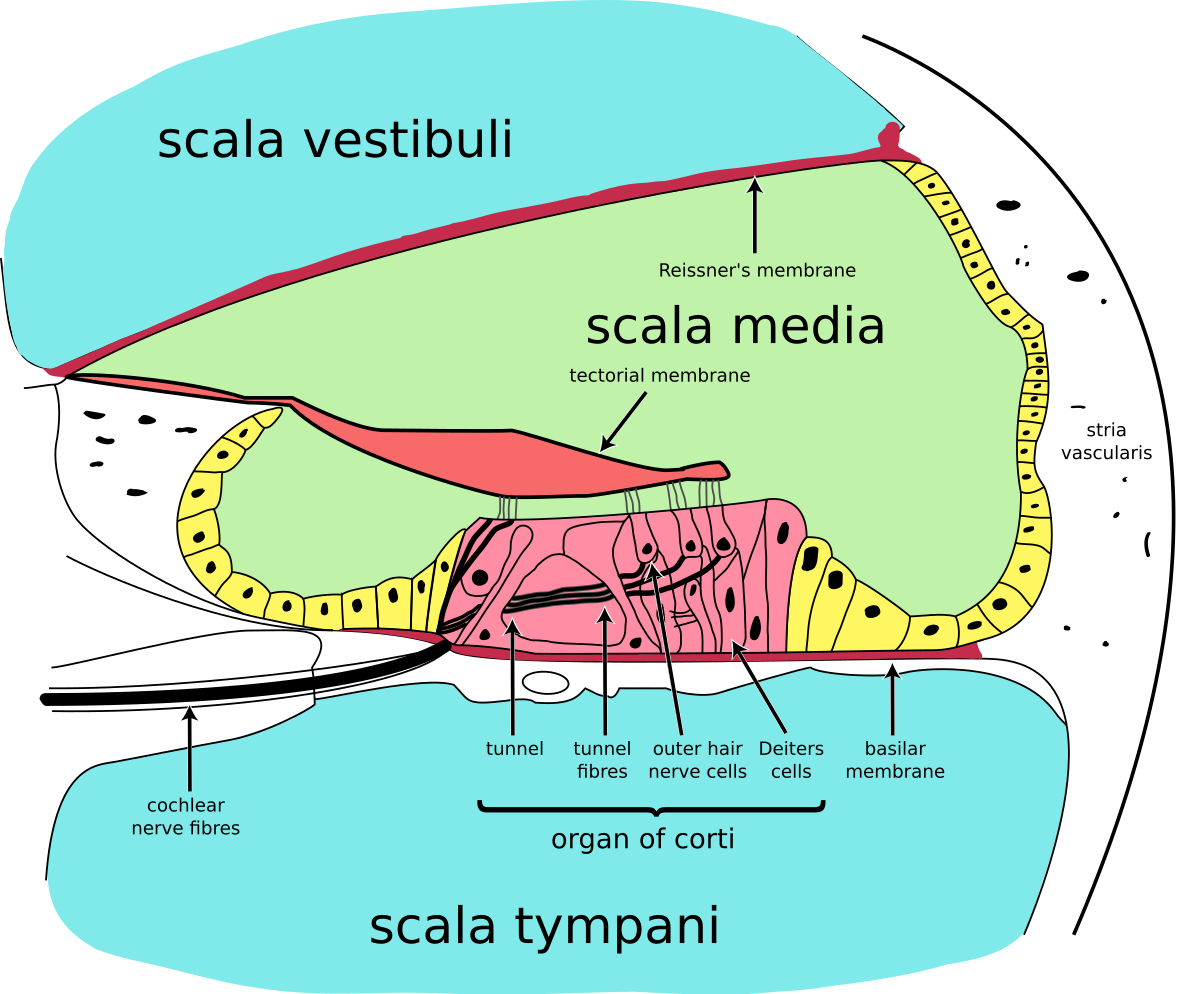
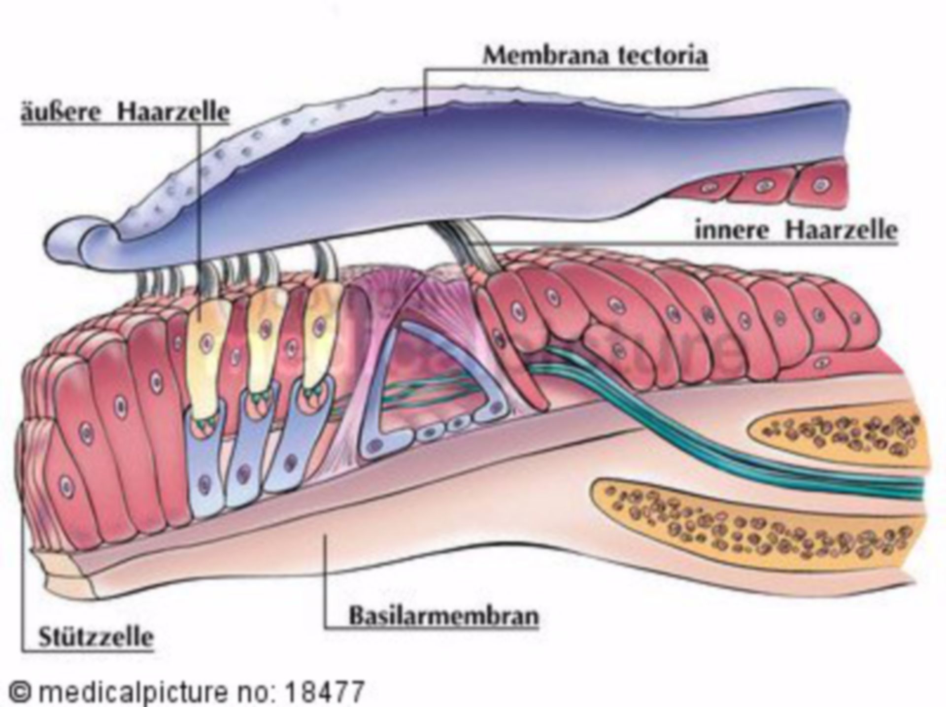
Diagram of Organ of Corti Cross-Section. Back to the Auditory System. Nolte (1993) The Human Brain 3rd Ed.Fig. 9-34B, p. 213. Cross-section through the Organ of Corti.. Start studying organ of corti. Learn vocabulary, terms, and more with flashcards, games, and other study tools.
L'organe de Corti est l'organe de la perception auditive. Il est constitué de cellules sensorielles (cellules ciliées internes ou CCI et cellules ciliées externes CCE) et de cellules de soutien (cellule de Deiters).

Organ of corti diagram
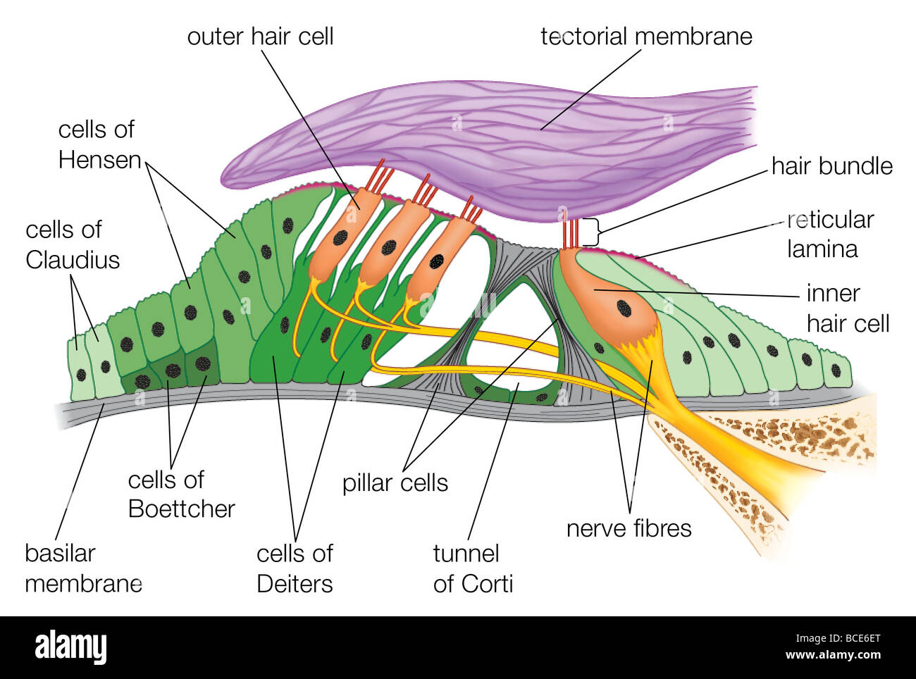
August 11, 2021 - Other articles where organ of Corti is discussed: human ear: Structure of the cochlea: …the basilar membrane is the organ of Corti, which contains the hair cells that give rise to nerve signals in response to sound vibrations. The side of the triangle is formed by two tissues that line the ... Download scientific diagram | Schematic representation of the organ of Corti. The figure shows the different cell types and extracellular structures in the organ of Corti. Abbreviations: TM (tectorial membrane), OHC (outer hair cells), IHC (inner hair cells), HB (hair bundle), SC (supporting ... How the Ear Works: Organ of Corti - The Temple of Hearing...
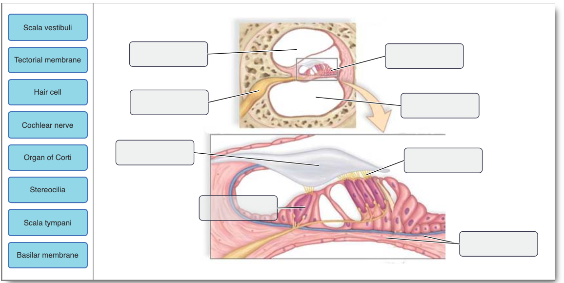
Organ of corti diagram. However, in the IHCs, the ‘W’ formation is wide and its long axis is linear and arranged at a right angle to the radial axis of the organ of Corti; also, the ciliary bundles are freestanding (with a few exceptions in the basal turn). This arrangement in the IHCs would be best suited for ... The organ of Corti is a specialized sensory epithelium that allows for the transduction of sound vibrations into neural signals. The organ of Corti itself is located on the basilar membrane. The organ of Corti rests on the basilar membrane and contains two types of hair cells: inner hair cells and outer hair cells. Inner hair cells transduce sound from vibrations to neural signals via the shearing action of their stereocilia. Diagram Of Organ Of Corti Nervous System Explore the Nerves with Interactive April 16th, 2019 - The spinal cord is a long thin mass of bundled neurons that carries information through the vertebral cavity of the spine beginning at the medulla oblongata of the brain on its superior end and continuing inferiorly to the Start studying Organ of Corti. Learn vocabulary, terms, and more with flashcards, games, and other study tools. Search. Create. Log in Sign up. Log in Sign up. 6 terms. begonia_blossom. Organ of Corti. STUDY. PLAY. Inner hair cell (IHC) Receptive Cell of the auditory apparatus. Single row of 3500 cells which are closer to the central axis of the coiled cochlea . Outer Hair …
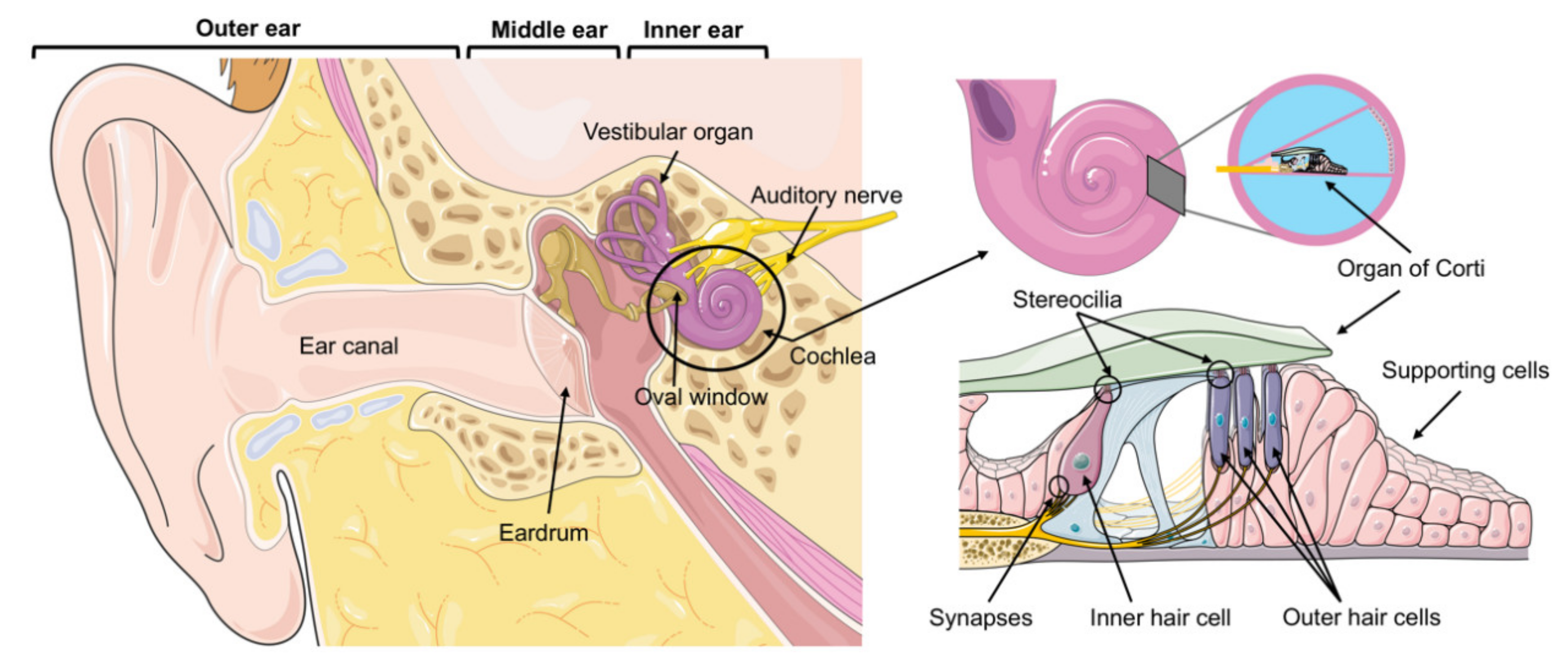
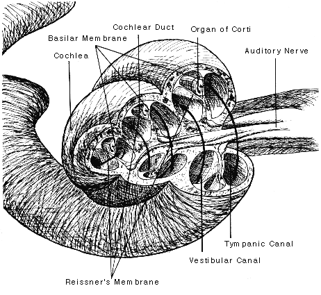
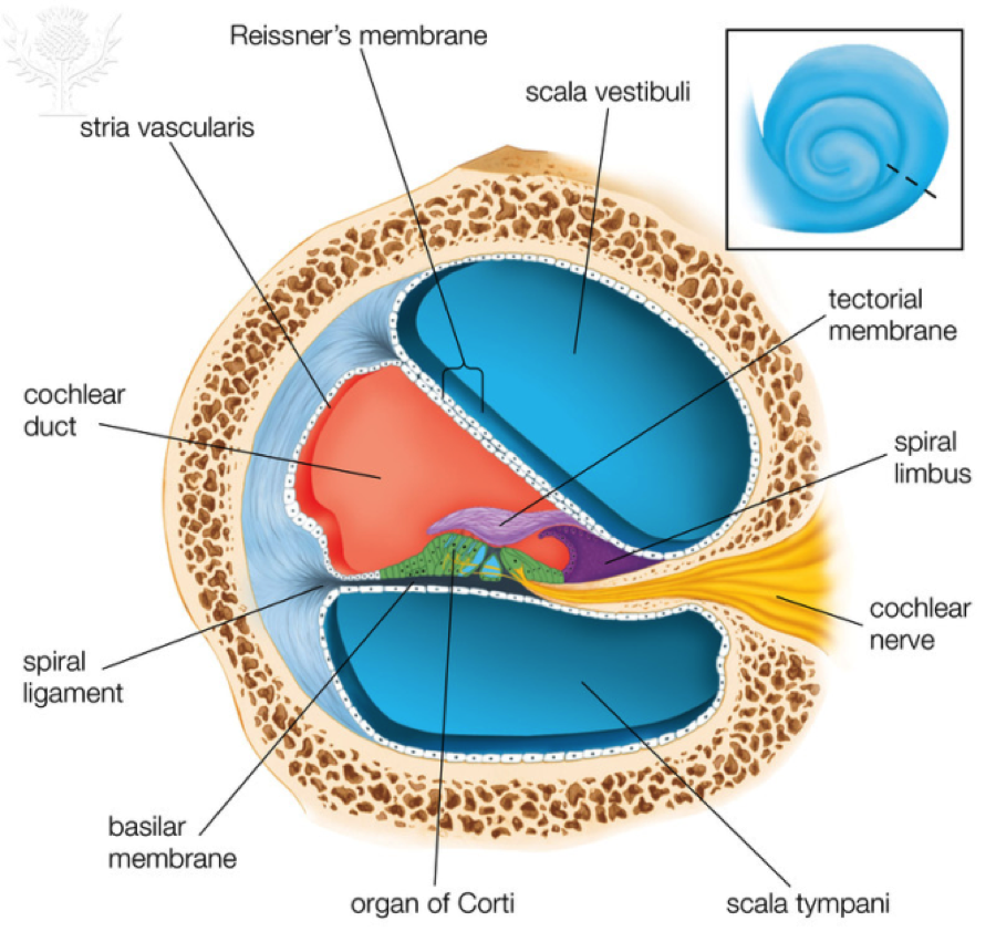
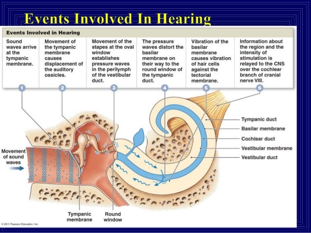
The cochlear duct is the triangular shaped section of the cochlea, which contains the organ of Corti. The oval window is quite simply an oval shaped window that is moved inwards by the movement of the stapes footplate. The scala vestibuli is the semicircle shaped region above the scala media and contains perilymph. Within the cochlea, a complex assemblage of hair cells, supporting cells, and membranes (tectorial and basilar membranes) constitutes the organ of Corti, now preferentially called the spiral organ. Three-dimensionally, this can be visualized as somewhat like a ribbon of tissue coiling upward from the base to the apex of the cochlea. Organ of Corti is located in middle cochlear canal in the inner ear. It has hair-like cells (mechanoreceptors) on its basilar membrane. When the fluid in the cochlea moves and vibrates due to the sound waves, the hair cells also vibrates. The Organ of Corti is an organ of the inner ear located within the cochlea which contributes to audition. The Organ of Corti includes three rows of outer hair cells and one row of inner hair cells. Vibrations caused by sound waves bend the stereocilia on these hair cells via an electromechanical force. The hair cells convert mechanical energy into electrical energy that is transmitted to the ...
February 2, 2014 - The Organ of Corti is a part of the cochlea and it mediates the sense of hearing transducing pressure waves to action potentials. This structure is localized in the scala media and it is formed by a series of hair cells, nervous terminations of spiral ganglion and supporting cells. Organ of Corti. Organ of Corti is the auditory organ of the cochlear duct, present on the basilar membrane, which separates scala tympani with scala media. Organ of Corti contains the mechanoreceptors of the ear in the form of hair cells. These hair cells are present in rows on the internal side of the organ of Corti that act as auditory receptors. 11. The organ of corti is a structure present in a. External ear b. Middle ear c. Semi circular canal d. Cochlea VERY SHORT ANSWER TYPE QUESTIONS 1. Rearrange the following in the correct order of involvement in electrical impulse movement-Synaptic knob, dendrites, cell body, Axon terminal, Axon 18-04-2018 Dec 18, 2020 · Diagram Of Organ Of Corti. Last Updated on Fri, 18 Dec 2020 | Unity Companies. The inner ear is housed in a maze of temporal bone passageways called the bony labyrinth, which is lined by a system of fleshy tubes called the membranous labyrinth (fig. 16.11). Between the bony and membranous labyrinths is a cushion of fluid called perilymph (PER-ih-limf), similar to cerebrospinal fluid.
The scala media is filled with endolymph and contains the auditory organ, the organ of Corti. Each organ of Corti contains ~18000 hair cells. Hair cells are present in the basilar membrane, which separates scala media from scala tympani. Stereocilia project from the hair cells and extend till the cochlear duct.
Jan 20, 2018 · The organ of Corti consists of supporting cells and many thousands of sensory hair cells. Each hair cell has up to 100 bristle-like hairs that translate mechanical movement into electrical sensory ...
Diagram Of Organ Of Corti BBC Science amp Nature Human Body and Mind Nervous September 23rd, 2014 - Ears nerves and brain Your ears are your organs of hearing In order to hear however you also need your cochlear nerves to transmit nerve impulses to your brain which then interpret the Cochlea Wikipedia April 15th, 2019 - The cochlea is filled
Diagram Of Organ Of Corti biology 105 anatomy amp physiology course online video, neuroscience for kids the ear, petrous part of temporal bone radiology reference, ear siumed edu, critical band wikipedia, human ear cross section images stock photos amp vectors, ear anatomy structure and parts of the ear information, chapter 15 test the special
The organ of Corti, or spiral organ, is the receptor organ for hearing and is located in the mammalian cochlea.This highly varied strip of epithelial cells allows for transduction of auditory signals into nerve impulses' action potential. Transduction occurs through vibrations of structures in the inner ear causing displacement of cochlear fluid and movement of hair cells at the organ of Corti ...
In this video, we explore the cochlea and the Organ of Corti by viewing it under light microscopy.
diagram slides for powerpoint is a collection of over 1000 impressively designed data driven chart and editable diagram s guaranteed to impress any audience, sensory coding loud vs soft sounds the organ of corti allows us to discriminate between different sound intensities loud sounds produce more
Title: Diagram Of Organ Of Corti Author: OpenSource Subject: Diagram Of Organ Of Corti Keywords: diagram of organ of corti, chapter 15 test the special senses flashcards quizlet, ear anatomy diagram amp pictures body maps healthline, pearson the biology place prentice hall, what funnels sound waves into the ear canal of the ear, biology 105 anatomy amp physiology course online video, ppt ...
Download scientific diagram | Micromechanics of the organ of Corti. (a) Two pillar cells (PC) form a stiff triangle on the basilar membrane; the liquid-filled interior space is referred to as ...
Diagram Of Organ Of Corti human ear cross section Images Stock Photos amp Vectors April 19th, 2019 - Find human ear cross section Stock Images in HD and millions of other royalty free stock photos illustrations and vectors in the Shutterstock collection Thousands of new high quality pictures added every day NervousSystemExploretheNerves with ...
May 8, 2018 - The mammalian hearing organ is a regular array of two types of hair cells surrounded by six types of supporting cells. Along the tonotopic axis, this conserved radial array of cell types shows longitudinal variations to enhance the tuning properties of basilar membrane.
The organ of Corti of the mammalian inner ear contains sensory hair cells and supporting cells in the auditory sensory epithelia. These cells are arranged to form a checkerboard-like cellular pattern. However, cellular and molecular mechanisms that produce this characteristic arrangement of cells had remained unknown for a long time.
Draw the diagram showing the internal structure of human ear and label the following parts. (a) Organ of Corti (b) Auditory nerve
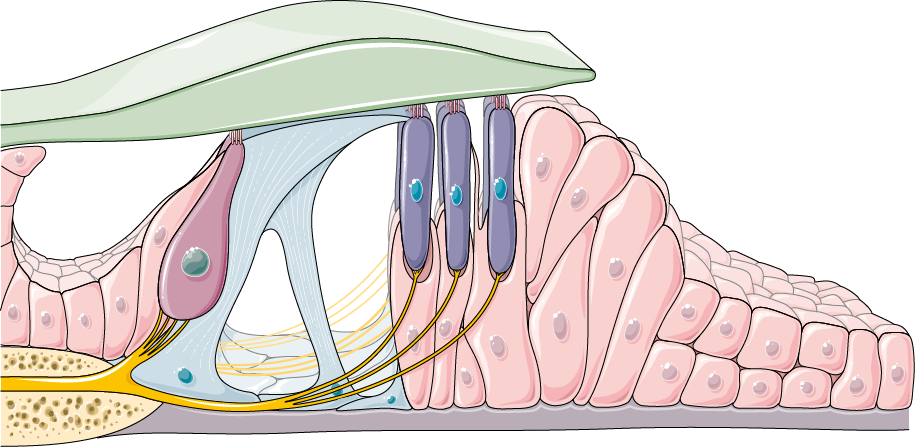
Schematic of the organ of Corti. In this transverse section of the basal part of a mammalian cochlea, 1 IHC (1) and 3 OHCs (2) are represented on either side of the tunnel of Corti (3). The tectorial membrane (6), floating in endolymph, caps the tallest stereocilia of the hair cells. The IHC is surrounded by supporting cells, whereas OHCs are ...
The meaning of ORGAN OF CORTI is a complex epithelial structure in the cochlea that contains thousands of hair cells, rests on the internal surface of the basilar membrane, and in mammals is the chief part of the ear by which sound waves are perceived and converted into nerve impulses to be ...
The organ of corti is a tube with hair-like cells that take the sound vibrations and changes them into electricity. Our brain runs on electricity; for it to understand sound, we have to change sound into electricity. When the hair-like cells are pushed by the sound, an important change happens. ...
August 11, 2021 - human ear - human ear - Organ of Corti: Arranged on the surface of the basilar membrane are orderly rows of the sensory hair cells, which generate nerve impulses in response to sound vibrations. Together with their supporting cells they form a complex neuroepithelium called the basilar papilla, ...
Jun 10, 2021 · Figure 10.21 A scanning electron micrograph of the hair cells of the spiral organ (organ of Corti). association of the basilar membrane, hair cells with sensory fibers, and tectorial membrane forms a functional unit called the spiral organ, or organ of Corti (fig. 10.22). When the cochlear duct is displaced by pressure waves of perilymph, a shearing force is created between the basilar membrane and the tectorial membrane.
Diagram Of Organ Of Corti What funnels sound waves into the ear canal of the ear April18th,2019 ...
Download scientific diagram | Components of the organ of Corti. ( A ) The organ of Corti rests on the basilar membrane, and is composed of the sensory receptor cells (OHCs and IHCs), supporting cells ( yellow ), and the tectorial membrane (TM). ( B ) The hair cell receptors are innervated by ...
http://www.interactive-biology.com - In this video, I talk about the organ of corti, which is found on top of the basiliar membrane inside the cochlea. When ...
Abstract. The cochlea resolves low-level acoustic signals into their individual frequency components and converts these into the auditory neural code. Vibration
Human Body. The Ear: Organs of Hearing and Balance Chart (d) 20x26. AS-IS: Obtain this 20" x 26" (51 x 66 cm) otolaryngological poster that illustrates ear anatomy, including right auricle, right tympanic membrane, middle ear, auditory ossicles, membranous labyrinth, membranous ampulla, organ of corti, macula of saccule.
The organ of Corti is the sensitive element in the inner ear and can be thought of as the body's microphone. It is situated on the basilar membrane in one of the three compartments of the Cochlea.It contains four rows of hair cells which protrude from its surface. Above them is the tectoral membrane which can move in response to pressure variations in the fluid- filled tympanic and vestibular ...
January 14, 2021 - Organ of Corti - the organ of Corti is the receptor organ for hearing. It rests on the surface of the basilar membrane in the cochlea and contains hair cells, which transduce vibrations caused by sound waves into electrical impulses that can be interpreted by the brain.
How the Ear Works: Organ of Corti - The Temple of Hearing...
Download scientific diagram | Schematic representation of the organ of Corti. The figure shows the different cell types and extracellular structures in the organ of Corti. Abbreviations: TM (tectorial membrane), OHC (outer hair cells), IHC (inner hair cells), HB (hair bundle), SC (supporting ...
August 11, 2021 - Other articles where organ of Corti is discussed: human ear: Structure of the cochlea: …the basilar membrane is the organ of Corti, which contains the hair cells that give rise to nerve signals in response to sound vibrations. The side of the triangle is formed by two tissues that line the ...










![Figure, Organ of Corti. A. Cross...] - StatPearls - NCBI ...](https://www.ncbi.nlm.nih.gov/books/NBK538335/bin/Figure__1A.jpg)
0 Response to "38 organ of corti diagram"
Post a Comment