40 Arteries Of The Brain Diagram
Main arteries of the body. | Body anatomy, Human body ... By the end of this section, you will be able to: Identify the vessels through which blood travels within the pulmonary circuit, beginning from the right ventricle of the heart and ending at the left atrium Create a flow chart showing the major systemic arteries through which blood travels from the aorta and its major branches, to the most significant arteries feeding into the right and left ... Head Arteries & Nerves Anatomy, Function & Diagram | Body Maps In the brain, a circle consisting to of two carotid arteries and the basilar artery form the circle of Willis. This supplies blood in the center of the brain and branches to the cerebrum, pons,...
7 Arteries of the brain [110]. | Download Scientific Diagram Download scientific diagram | 7 Arteries of the brain [110]. from publication: Computational Models of Cerebral Hemodynamics | The cerebral tissue requires a constant supply of oxygen and nutrients.
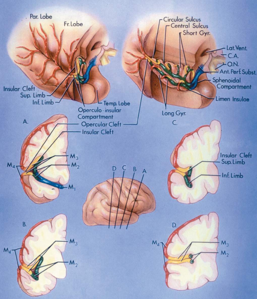
Arteries of the brain diagram
Coronary Arteries: How It Works & Images - Cleveland Clinic Coronary Arteries. The heart receives its own supply of blood from the coronary arteries. Two major coronary arteries branch off from the aorta near the point where the aorta and the left ventricle meet. These arteries and their branches supply all parts of the heart muscle with blood. Brain Circulation - University of Massachusetts Medical School Brain Circulation. The brain derives its arterial supply from the paired carotid and vertebral arteries. Every minute, about 600-700 ml of blood flow through the carotid arteries and their branches while about 100-200 ml flow through the vertebral-basilar system. The carotid and vertebral arteries begin extracranially, and course through the ... Neuroanatomy, Brain Arteries Article Introduction. The brain receives vascular supply from a network of arteries that anastomose to form the circle of Willis. Because the brain has a constant high metabolic demand and no energy supply of its own, it requires a significant blood supply, consuming 15% of total cardiac output, and any blockage of blood flow leads to severe damage and a host of neurological pathologies.
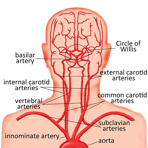
Arteries of the brain diagram. The Arteries (Human Anatomy): Picture, Definition ... The arteries are the blood vessels that deliver oxygen-rich blood from the heart to the tissues of the body. Each artery is a muscular tube lined by smooth tissue and has three layers: The intima ... Vascular anatomy - NeurologyNeeds.com The brain is supplied by branches of the internal carotid artery anteriorly and by branches of the vertebral artery posteriorly. The aortic arch gives off three great vessels: the brachiocephalic artery, the left common carotid artery and the left subclavian artery. Brain Blood Supply: Anatomy & Diagrams - Video & Lesson ... The main arteries supplying your brain include the internal carotid artery, which is a branch of the common carotid artery that supplies your brain with oxygenated blood; the anterior cerebral ... Anatomical diagrams of the brain - eAnatomy The study of the arterial supply of blood to the brain is facilitated by a diagram showing the cerebral arterial vascular areas in lateral and medial views and axial and coronal section and by diagrams of arteries forming the Willis' circle (internal and vertebral carotid arteries, basilar artery, anterior and posterior communicating arteries ...
Coronary Artery Diagramming - Resus Review The normal major epicardial arteries are shown in the figure below. The left coronary cusp gives rise to the left main coronary artery which branches to the left anterior descending (LAD) and left circumflex artery (LCx, Circ). The branches from the LAD are labelled in sequence diagonal 1, diagonal 2, etc. Midsagittal Arteries of the Brain - SmartDraw Midsagittal Arteries of the Brain. Create healthcare diagrams like this example called Midsagittal Arteries of the Brain in minutes with SmartDraw. SmartDraw includes 1000s of professional healthcare and anatomy chart templates that you can modify and make your own. Major arteries, veins and nerves of the body: Anatomy | Kenhub Arteries, veins and nerves of the trunk (diagram) Arteries of the trunk include the: thoracic aorta, celiac trunk, superior mesenteric artery, inferior mesenteric artery, and common iliac arteries (with its terminal branches internal iliac and external iliac arteries ). Upper extremity Arteries, veins and nerves of the arm (a diagram) Arteries of the Brain Diagram | Quizlet Start studying Arteries of the Brain. Learn vocabulary, terms, and more with flashcards, games, and other study tools.
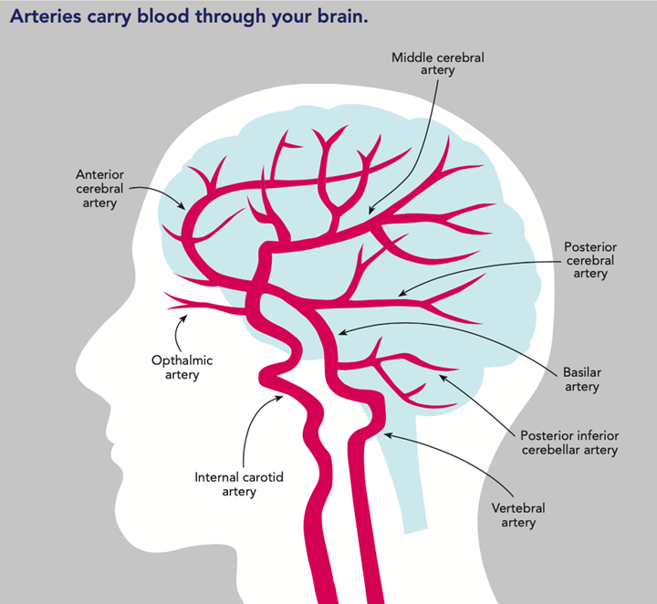
Veins Of The Human Body Diagram - Studying Diagrams Nov 26 2020 - This diagram shows the major veins in the human body. A medical diagram showing the heart arteries and veins of the human body. Avg turnaround time to acceptance for papers submitted to GE Collections is 102 days. Coronary Intervention - First Coast Heart Vascular Center. They are thick elastic and are divided into a small. › BrainstemBrainstem - Physiopedia The brain stem receives its blood supply exclusively from the posterior circulation, including the vertebrae and basilar artery. The medulla receives its blood supply from the vertebral via medial and lateral perforating arteries. The pons and midbrain receive their blood from the basilar via the medial and lateral perforating arteries. Major Arteries of the Head and Neck - TeachMeAnatomy Other Arteries of the Neck. The neck is supplied by arteries other than the carotids. The right and left subclavian arteries give rise to the thyrocervical trunk. From this trunk, several vessels arise, which go on to supply the neck. The first branch of the thyrocervical trunk is the inferior thyroid artery. Neuroanatomy, Brain Arteries - StatPearls - NCBI Bookshelf The brain is supplied by the internal carotid arteries (ICA), which branch from the common carotid arteries, and the vertebral arteries, which branch from the subclavian. The ICA gives rise to the anterior cerebral artery (ACA) and the middle cerebral artery (MCA).
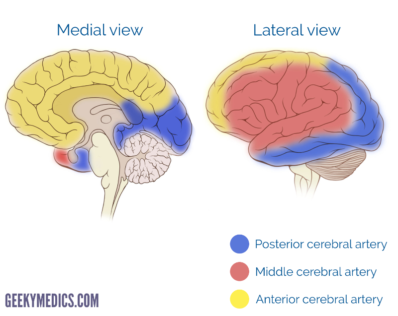
PDF Arteries of the Head and Neck - Exploring Nature They enter the skull through the carotid canalsand branch into: > ophthalmic arterieswhich supply blood to the eye sockets, anterior scalp and nasal cavity > anterior cerebral arterieswhich supply blood to the medial cerebral hemispheres > middle cerebral arterieswhich supply blood to the lateral temporal lobes and parietal lobes.
courses.lumenlearning.com › chapter › arteriesArteries | Boundless Anatomy and Physiology Arteries are blood vessels that carry blood away from the heart. This blood is normally oxygenated, with the exception of blood in the pulmonary artery. Arteries typically have a thicker tunica media than veins, containing more smooth muscle cells and elastic tissue. This allows for modulation of vessel caliber and thus control of blood pressure.

Vintage anatomy print of the human brain depicting the nerves and arteries. Poster Print by John Parrot/Stocktrek Images - Item # VARPSTJPA700059H
› human-brain-functionsHow Human Brain Functions? Brain Functioning List with Diagram It keeps the diastolic (minimum) and systolic (maximum) pressure in the arteries under normal limits. In the case the blood pressure rises beyond the bearable limits, you are very likely to suffer from a heartattack, brain haemorrhage or other critical circulatory disorders.
anatomy.co.uk › aortaThe Aorta - Anatomy, Location, Structure, Function & Diagram The main artery of the body begins at the heart and ends when it divides itself in the iliac arteries to supply blood to the lower limbs. The aorta is found in the trunk of the human body and it is divided and named according to its location in thoracic aorta and abdominal aorta.
Carotid Artery (Human Anatomy): Picture, Definition ... The internal carotid artery supplies blood to the brain. The external carotid artery supplies blood to the face and neck. Like all arteries, the carotid arteries are made of three layers of tissue:
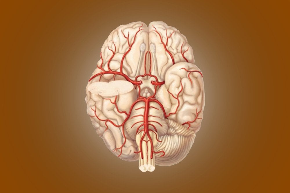
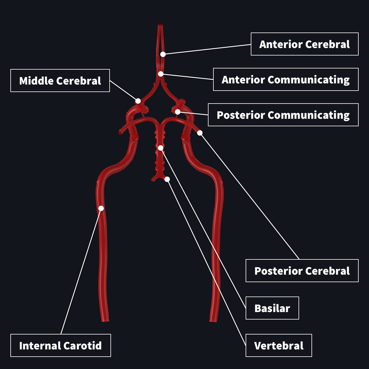
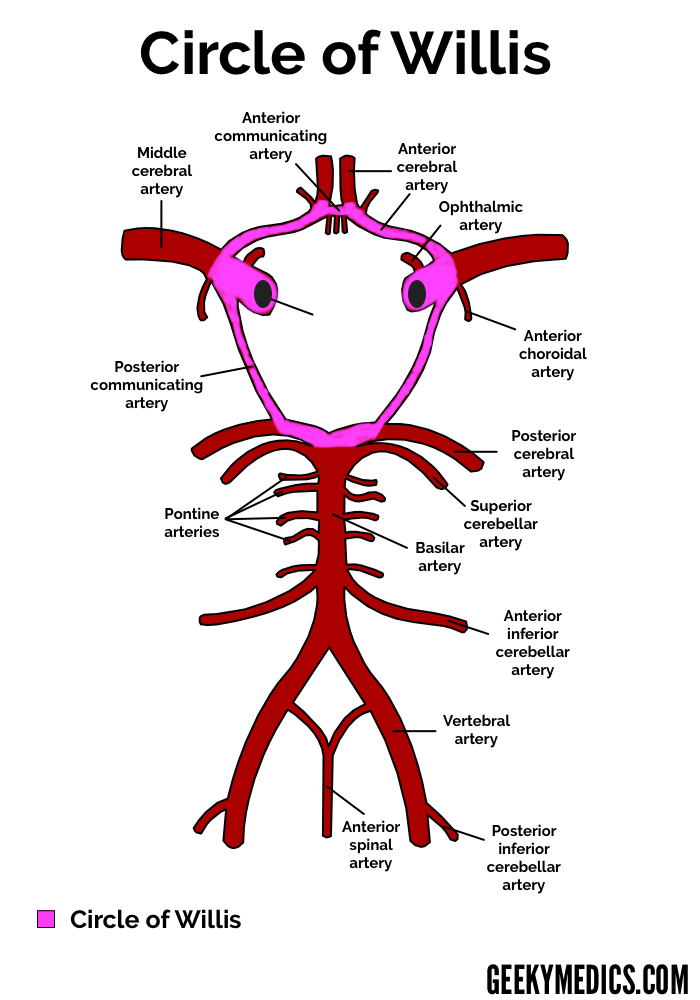
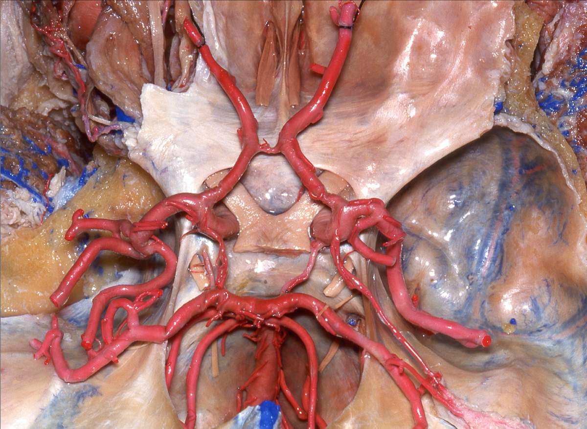
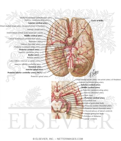
Blood supply to the brain: Anatomy of cerebral arteries ... Arteries of the brain and 'circle of Willis' diagram There is a point at which the anterior and posterior arterial circuits of the brain unite or anastomose. This area is known as the circle of Willis. It is a central communication that unites the internal carotid and vertebrobasilar systems. Circle of Willis is indeed a hot neuroanatomy topic!
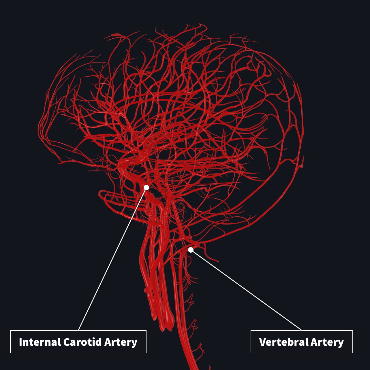
› books › NBK11042The Blood Supply of the Brain and Spinal Cord - Neuroscience ... The brain receives blood from two sources: the internal carotid arteries, which arise at the point in the neck where the common carotid arteries bifurcate, and the vertebral arteries (Figure 1.20).The internal carotid arteries branch to form two major cerebral arteries, the anterior and middle cerebral arteries.The right and left vertebral arteries come together at the level of the pons on the ...
Blood Supply To The Brain Diagram - Studying Diagrams The common carotid arteries supply blood to the head and neck. Label the diagram below Figure 31 The. This can be located in the. The brains blood supply keeps it hydrated which is critical since the brain is over 70 water. Blood brings the brain the hormones and neurotransmitters it needs to function.
› anatomy-of-the-brainBrain Anatomy and How the Brain Works - Hopkins Medicine The vertebral arteries follow the spinal column into the skull, where they join together at the brainstem and form the basilar artery, which supplies blood to the rear portions of the brain. The circle of Willis , a loop of blood vessels near the bottom of the brain that connects major arteries, circulates blood from the front of the brain to ...
PDF Blood Vessels of the Brain - UNC School of Medicine lood in the brain is supplied by two pairs of large blood vessels (arteries): the carotid arteries and the vertebral arteries: Carotid Arteries: These vessels run along the front of the neck. There is a right-sided carotid and a left-sided carotid artery. If a stroke happens in this area, it can cause changes with speech, vision and sensation.
Basilar Artery | Brain Parts The diagram shows the main branches of the basilar artery. The basilar artery is formed by the two vertebral arteries and travel as a single artery over the upper medulla and the entire pons. Its terminal division is into the right and left posterior cerebral arteries. Its first branch after this division is the posterior communicating artery.
VI. The Arteries. 3b. The Arteries of the Brain. Gray ... The parts of the brain included within this arterial circle are the lamina terminalis, the optic chiasma, the infundibulum, the tuber cinereum, the corpora mammillaria, and the posterior perforated substance. 2: FIG. 519- Diagram of the arterial circulation at the base of the brain. A.L. Antero-lateral. A.M. Antero-medial. P.L. Postero-lateral.
Blood supply to the brain | Complete Anatomy - 3D4Medical The basilar artery ends by bifurcating into posterior cerebral arteries . Every time the heart beats, arteries carry 20 to 25% of the blood to the brain. The blood flow to the brain is vital to its function since it is particularly sensitive to oxygen starvation. When an area of the brain is cut off from blood flow, a stroke can result.
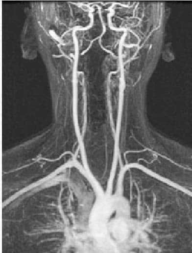
Arterial Supply to the Brain - Carotid - Vertebral ... Fig 1.0 - Arteriogram of the arterial supply to the CNS. There are two paired arteries which are responsible for the blood supply to the brain; the vertebral arteries, and the internal carotid arteries. These arteries arise in the neck, and ascend to the cranium.
Arteries of the Brain - Brain Basics The brain isn't a vascular organ, but blood is a very important necessity for it. Blood carry's nutrients to the brain tissues and keep the brain healthy. When blood flow stops, the brain suffers a stroke, damaging it. The brain is filled with arteries and segments that branch out over all the lobes.
The Arteries of the Brain - Human Anatomy FIG.519- Diagram of the arterial circulation at the base of the brain.A.L. Antero-lateral.A.M. Antero-medial.P.L. Postero-lateral.P.M. Posteromedial ganglionic branches. Since the mode of distribution of the vessels of the brain has an important bearing upon a considerable number of the pathological lesions which may occur in this part of the nervous system, it is important to consider a ...
› human-body-maps › leg-vesselsLeg Vessels Anatomy, Function & Diagram | Body Maps Jan 22, 2018 · A web of blood vessels—arteries, veins, and capillaries—circulate blood to organs, muscles, bones, and other tissues. ... The frontal lobe is the part of the brain that controls important ...
Neuroanatomy, Brain Arteries Article Introduction. The brain receives vascular supply from a network of arteries that anastomose to form the circle of Willis. Because the brain has a constant high metabolic demand and no energy supply of its own, it requires a significant blood supply, consuming 15% of total cardiac output, and any blockage of blood flow leads to severe damage and a host of neurological pathologies.
Brain Circulation - University of Massachusetts Medical School Brain Circulation. The brain derives its arterial supply from the paired carotid and vertebral arteries. Every minute, about 600-700 ml of blood flow through the carotid arteries and their branches while about 100-200 ml flow through the vertebral-basilar system. The carotid and vertebral arteries begin extracranially, and course through the ...
Coronary Arteries: How It Works & Images - Cleveland Clinic Coronary Arteries. The heart receives its own supply of blood from the coronary arteries. Two major coronary arteries branch off from the aorta near the point where the aorta and the left ventricle meet. These arteries and their branches supply all parts of the heart muscle with blood.

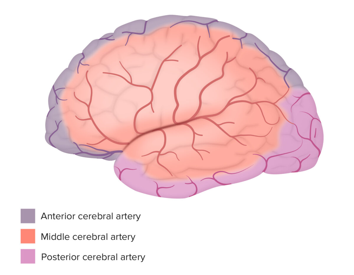


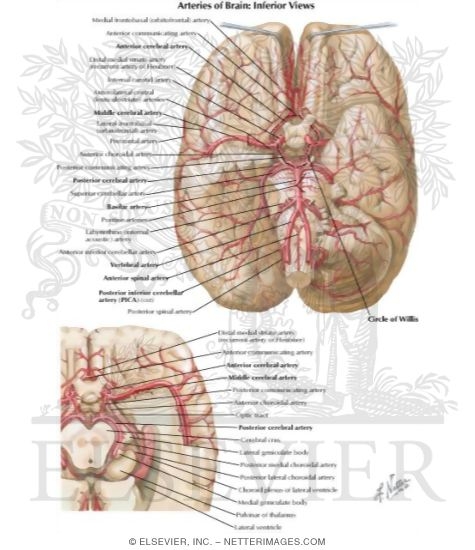
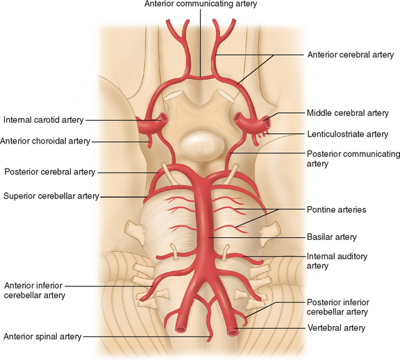






/CircleofWillis-87378170-3ece0502a02949dd82310d723e0d4c98.jpg)

:watermark(/images/watermark_only.png,0,0,0):watermark(/images/logo_url.png,-10,-10,0):format(jpeg)/images/anatomy_term/arteria-temporalis-media-2/ZJMcP8xJB97dDXsQ8d1R0Q_arteria-temporalis-media-2.jpg)

:background_color(FFFFFF):format(jpeg)/images/library/12166/arteries-of-brain-inferior-view_english.jpg)


:watermark(/images/watermark_only.png,0,0,0):watermark(/images/logo_url.png,-10,-10,0):format(jpeg)/images/anatomy_term/circulus-arteriosus-cerebri/qNRQ5TN7dGARxv8Qr5ysNg_Circulus_arteriosus_cerebri_01.png)


0 Response to "40 Arteries Of The Brain Diagram"
Post a Comment