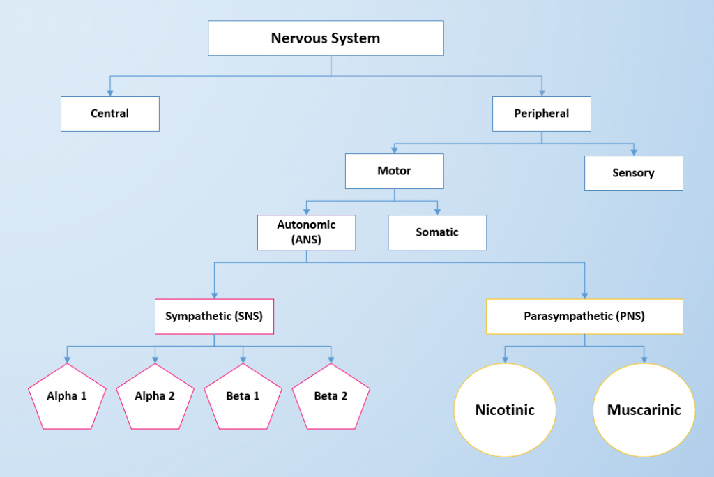
39 drag the labels onto the diagram to identify the divisions and receptors of the nervous system.
A&P 2 Lab 5 HW Flashcards | Quizlet Drag the labels onto the diagram to identify the components of the autonomic nervous system. ... Drag the labels to identify the anatomical differences between the sympathetic and parasympathetic divisions. PDF The Nervous System The Nervous System Functions of the Nervous System 1. Gathers information from both inside and outside the body - Sensory Function 2. Transmits information to the processing areas of the brain and spine 3. Processes the information in the brain and spine - Integration Function 4.
Answered: Drag the labels onto the diagram to… | bartleby Transcribed Image Text: < > A session.masteringaandp.com Content e MasteringAandP: Chapte pter 8 Quiz: Overview of the Skeleton - Classification and Structure of Bones and Cartilages - Attempt 1 cise 8 Review Sheet Art-labeling Activity 2 Part A Drag the labels onto the diagram to identify the structures of an osteon. Reset Help canaliculi central canal lamella lacuna Submit Request Answer ...
Drag the labels onto the diagram to identify the divisions and receptors of the nervous system.
PDF The Autonomic Nervous System and Visceral Sensory Neurons nervous system more slowly than through the somatic motor system. It is important to emphasize that the autonomic ganglia are motor ganglia containing the cell bodies of motor neurons, in contrast to the dorsal root ganglia, which are sensory ganglia. Divisions of the Autonomic Nervous System The ANS has two divisions, the sympathetic and parasym- 16.1 Divisions of the Autonomic Nervous System - Anatomy ... The autonomic nervous system regulates many of the internal organs through a balance of two aspects, or divisions. In addition to the endocrine system, the autonomic nervous system is instrumental in homeostatic mechanisms in the body. The two divisions of the autonomic nervous system are the sympathetic division and the parasympathetic division. Solved Drag the labels onto the diagram to identify the ... Transcribed image text: Prag the labels onto the diagram to identify the components of the autonomic nervous system! Reset Help Cardiac muscle Smooth muscle Brain Ganglionic neurons Preganglionic neuron Visceral Effectors Adipocytes Autonomic nuclei in spinal cord Autonomic nuclei in brain stem Spinal cord Autonomic ganglia Visceral motor nuclei in hypothalamus Glands Preganglionic neuron ...
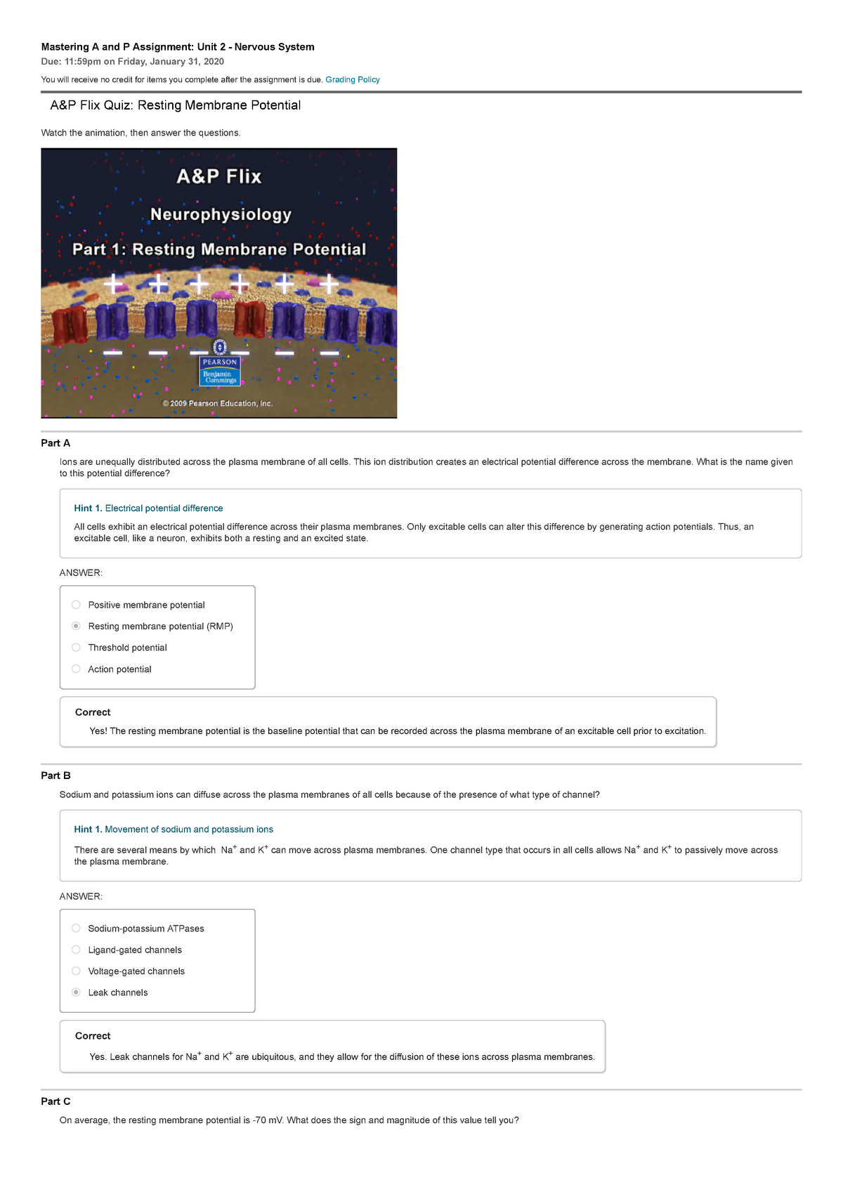
Drag the labels onto the diagram to identify the divisions and receptors of the nervous system.. 1.4 Anatomical Terminology - Anatomy & Physiology Figure 1.4.1 - Regions of the Human Body: The human body is shown in anatomical position in an (a) anterior view and a (b) posterior view. The regions of the body are labeled in boldface. A body that is lying down is described as either prone or supine. Major Body Cavities & their subdivisions Flashcards by ... The more inferior of the two enclose body cavities that protects the fragile nervous system organs; has two subdivisions, the cranial cavity and the vertebral cavity, which are continuous with each other ... contain tiny bones that transmit sound vibrations to the hearing receptors in the inner ears Decks in Anatomy & Physiology 1 Class (18 ... Drag the labels onto the diagram to identify the | Chegg.com Transcribed image text: Drag the labels onto the diagram to identify the divisions and receptors of the nervous system. Reset Help Organization of the Nervous System Integrate, process, and coordinate sensory data and motor commands cord) sensory Sensory information within afferent division Motor commands within efferent division Nervous includes neural tissue outside the CNS) Nervous sensory ... Part A Drag the labels onto the diagram to identify the ... Drag the labels to identify depolarization, repolarization, and hyperpolariztion. ANSWER: Correct A&P Flix Activity: Resting Membrane Potential Watch the A&P Flix Resting Membrane Potential video and then complete the activities below. Part A - Separation of Charges Across the Membrane of a Resting Neuron A resting neuron is an unstimulated neuron that is not presently generating an action ...
Divisions of the Autonomic Nervous System | Anatomy and ... The autonomic nervous system regulates many of the internal organs through a balance of two aspects, or divisions. In ition to the endocrine system, the autonomic nervous system is instrumental in homeostatic mechanisms in the body. The two divisions of the autonomic nervous system are the sympathetic division and the parasympathetic division. Chapter 11 Homework Flashcards | Quizlet Drag the appropriate labels to their respective targets. ... Which of the cell types shown helps determine capillary permeability in the CNS? A&P2 Lab 7 HW, A&P2 Lab 6 HW, A&P 2 Lab 5 HW ... - Quizlet Drag the labels onto the diagram to identify the pituitary hormones that ... and the letter ______ represents the efferent division of the nervous system. Chap 16 Autonomic Nervous System Flashcards - Quizlet Drag the labels onto the diagram to identify the components of the autonomic nervous system. ... Which anatomical description is true of the parasympathetic division of the autonomic nervous system? ... activates nicotinic receptors in the peripheral nervous system. This means it will _____.
PDF Palm Beach State College Palm Beach State College Mastering A and P Assignment Unit 2 - Nervous System ... Drag the labels onto the diagram of neurochemical communication at an autonomic synapse. ANSWER: Correct. Art-labeling Activity Figure 11. Label the parts of the neuromuscular junction. Part A. Drag the labels onto the diagram to identify parts of the neuromuscular junction. ANSWER: Reset Help. ... Efferent Divisions of the Nervous System. receptor and effector diagram burton chaseview overall receptor and effector diagram. finger tracing calm down cards receptor and effector diagram. By hydralazine and isosorbide dinitrate trade name; No Comments 10.2 Skeletal Muscle - Anatomy & Physiology Figure 10.2.2 - Muscle Fiber: A skeletal muscle fiber is surrounded by a plasma membrane called the sarcolemma, which contains sarcoplasm, the cytoplasm of muscle cells. A muscle fiber is composed of many myofibrils, which contain sarcomeres with light and dark regions that give the cell its striated appearance.
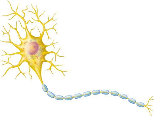
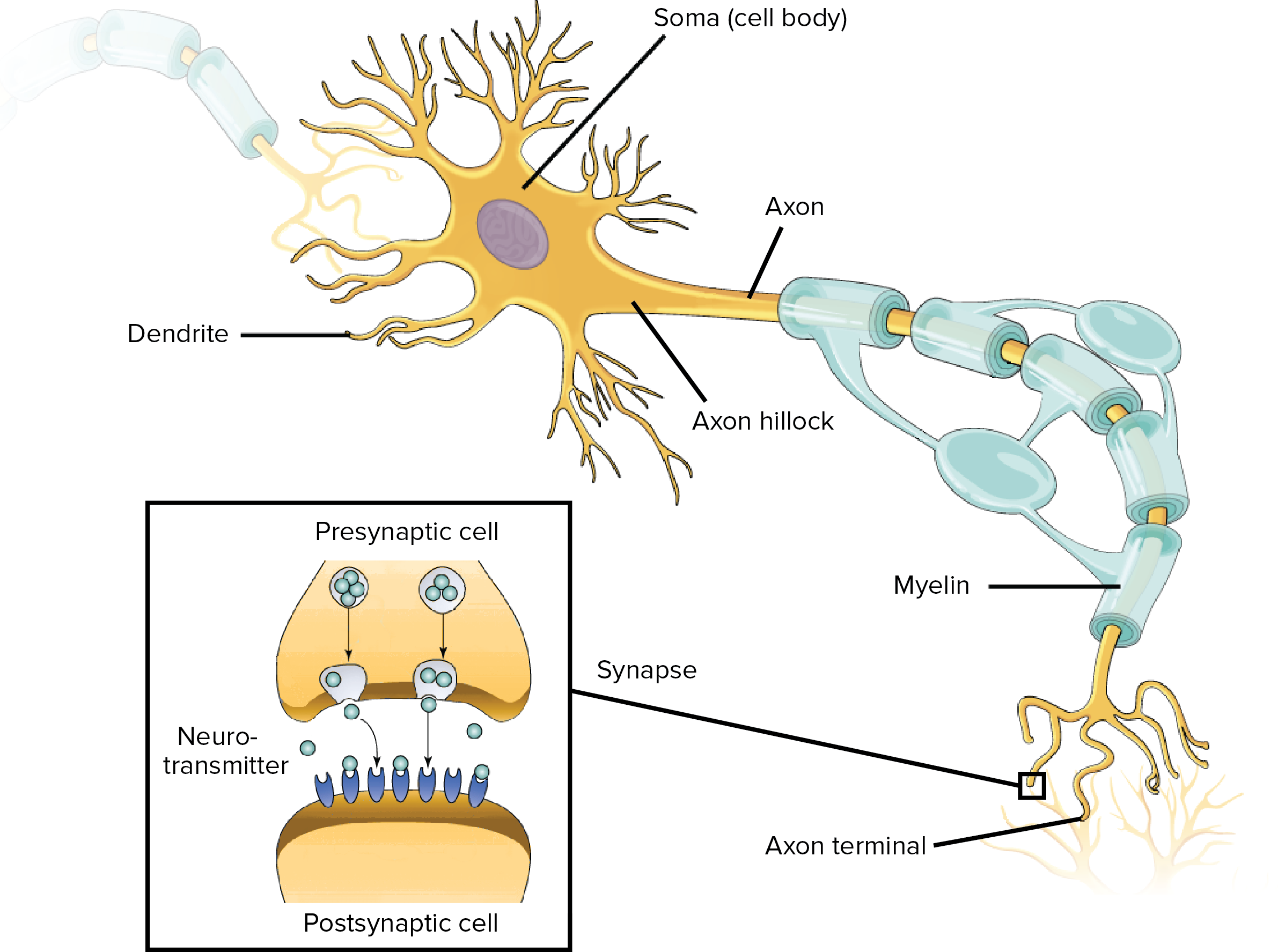
A&P2 Lab and Powerpoints Flashcards - Quizlet Drag the labels onto the diagram to identify the divisions and receptors of the nervous system. look at pic Drag the labels to identify the structural components of a typical neuron.
Solved Drag the labels onto the diagram to identify the ... View the full answer. Transcribed image text: Part A Drag the labels onto the diagram to identify the components of the somatic nervous system. Reset Help Brain Somatic motor nuclei of brain stem Somatic motor nuclei of spinal cord Spinal cord Skeletal muscle Upper motor neurons in primary motor cortex Lower motor neurons Submit Request Answer.
Drag The Labels Onto The Diagram To Identify Structural ... Drag the labels onto the diagram to identify the divisions and receptors of the nervous system. look at pic drag the labels to identify the structural components of a typical neuron. Drag the labels onto the diagram to identify the components of the integumentary system. look at pic the dermis is composed of the papillary layer and the .
A&P2 Lab 13 HW, A&P2 Lab 12 HW, A&P2 Lab 11 HW, A ... - Quizlet Drag the labels onto the diagram to identify the divisions and receptors of the nervous system. look at pic Drag the labels to identify the structural components of a typical neuron.
Neuroglia | Boundless Anatomy and Physiology Identify the neuroglia of the peripheral nervous system. Key Takeaways Key Points. There are two kinds of neuroglia in the peripheral nervous system (PNS): Schwann cells and satellite cells. Schwann cells provide myelination to peripheral neurons. Functionally, the schwann cells are similar to oligodendrocytes of the central nervous system (CNS).
PDF Chapter 12 Central Nervous System Copyright © 2010 Pearson Education, Inc. Figure 12.8a Functional and structural areas of the cerebral cortex. Gustatory cortex (in insula) Primary motor cortex
HW 7.pdf - HW 7 Due: 11:59pm on Friday, October 27, 2017 ... HW 7 Due: 11:59pm on Friday, October 27, 2017 To understand how points are awarded, read the Grading Policy for this assignment. Art-labeling Activity: Organization of the Nervous System Learning Goal: To learn the divisions and receptors of the nervous system. Label the divisions and receptors of the nervous system. Part A Drag the labels onto the diagram to identify the divisions and ...
A&P Chapter 11 Nervous System 2 Homework - Quizlet Drag and drop each label into the appropriate box, identifying which division of the autonomic nervous system is responsible for the given function.
14.5 Sensory and Motor Pathways - Anatomy & Physiology In the somatic nervous system, the thalamus is an important relay for communication between the cerebrum and the rest of the nervous system. The hypothalamus has both somatic and autonomic functions. In addition, the hypothalamus communicates with the limbic system, which controls emotions and memory functions.
A&P2 Lab 1 HW Flashcards - Quizlet Drag the labels onto the diagram to identify the divisions and receptors of the nervous system. look at pic Drag the labels to identify the structural components of a typical neuron.
Chap 15 Sensory Pathways and Somatic Nervous Sys. - Quizlet If a nerve impulse was transmitted to the central nervous system (CNS) on a C fiber, ... Drag the labels onto the diagram to identify the parts of the ...
A&P II Chapter 16 reading - Subjecto.com Drag the labels onto the figure to create a flow chart of how insulin and glucagon release change in different circumstances to keep blood glucose within a normal range. Look at picture. You are working in the free clinic when Father X comes in.
Solved Drag the labels onto the diagram to identify the ... This problem has been solved! See the answer. See the answer See the answer done loading. Drag the labels onto the diagram to identify the divisions and receptors of the nervous system. Show transcribed image text.
Solved Drag the labels onto the diagram to identify the ... Transcribed image text: Prag the labels onto the diagram to identify the components of the autonomic nervous system! Reset Help Cardiac muscle Smooth muscle Brain Ganglionic neurons Preganglionic neuron Visceral Effectors Adipocytes Autonomic nuclei in spinal cord Autonomic nuclei in brain stem Spinal cord Autonomic ganglia Visceral motor nuclei in hypothalamus Glands Preganglionic neuron ...
16.1 Divisions of the Autonomic Nervous System - Anatomy ... The autonomic nervous system regulates many of the internal organs through a balance of two aspects, or divisions. In addition to the endocrine system, the autonomic nervous system is instrumental in homeostatic mechanisms in the body. The two divisions of the autonomic nervous system are the sympathetic division and the parasympathetic division.
PDF The Autonomic Nervous System and Visceral Sensory Neurons nervous system more slowly than through the somatic motor system. It is important to emphasize that the autonomic ganglia are motor ganglia containing the cell bodies of motor neurons, in contrast to the dorsal root ganglia, which are sensory ganglia. Divisions of the Autonomic Nervous System The ANS has two divisions, the sympathetic and parasym-











0 Response to "39 drag the labels onto the diagram to identify the divisions and receptors of the nervous system."
Post a Comment