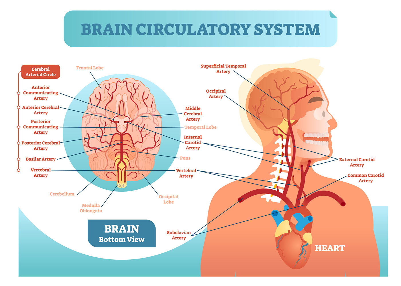
37 eye to brain connection diagram
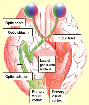
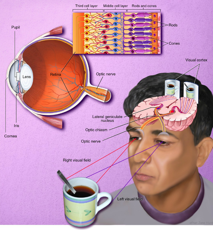
Studying Diagrams - Page 2 of 1210 - Study for utilizing ... Studying Diagrams. Study for utilizing diagrams are when you need to understand either a diagram process or a collection of connections How the Eye and the Brain Work Together - AbilityPath June 8, 2020 - The optic nerve of each eye meets the other at the optic chiasm. Medial nerves of each optic nerve cross, but lateral nerves stay on the same side. The overlap of nerve fibers allows for depth perception. Electrical impulses are communicated to the visual cortex of the brain by way of the optic ...
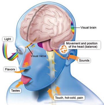
Retina - Wikipedia The retina (from Latin: rete "net") is the innermost, light-sensitive layer of tissue of the eye of most vertebrates and some molluscs.The optics of the eye create a focused two-dimensional image of the visual world on the retina, which translates that image into electrical neural impulses to the brain to create visual perception.

Eye to brain connection diagram
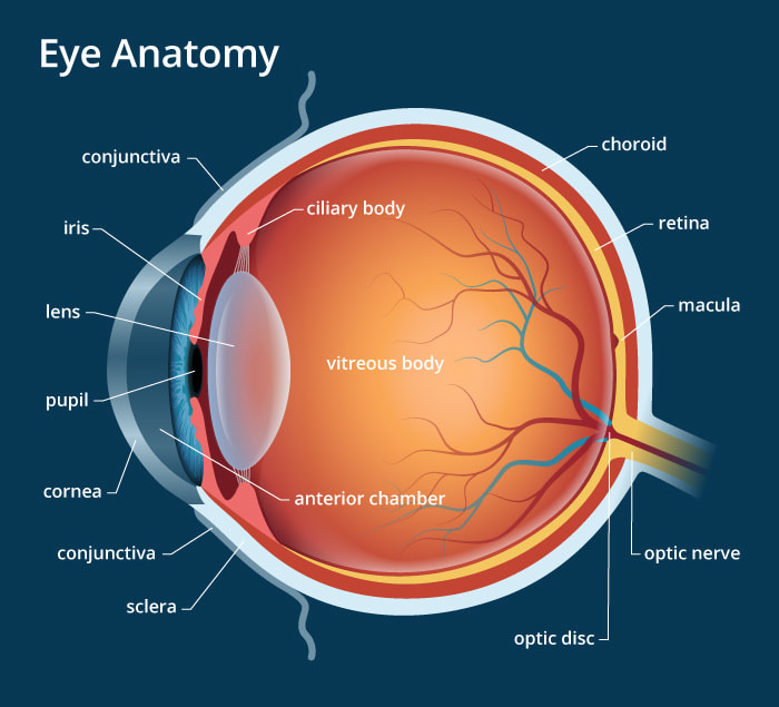
Our Sense of Sight: Part 1. Eye Anatomy and Function Eye Anatomy and Function · FEATURING: A "CLASS EXPERIMENT" PLUS: "TRY YOUR OWN EXPERIMENT" A Wiring Diagram of the Brain - MIT Technology Review Only one organism's wiring diagram currently exists: that of the microscopic worm C. elegans. Despite containing a mere 302 neurons, the C. elegans mapping effort took more than a decade to... Human eye - Wikipedia The approximate field of view of an individual human eye (measured from the fixation point, i.e., the point at which one's gaze is directed) varies by facial anatomy, but is typically 30° superior (up, limited by the brow), 45° nasal (limited by the nose), 70° inferior (down), and 100° temporal (towards the temple).
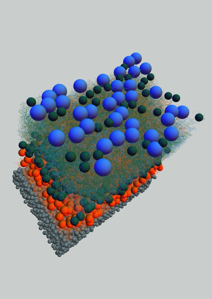
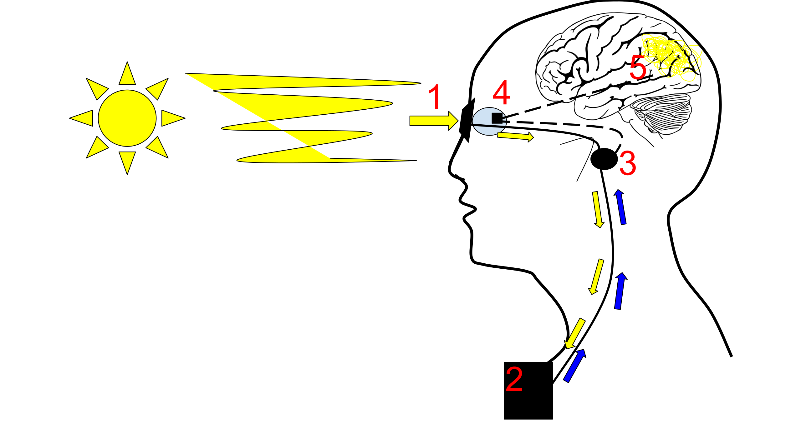
Eye to brain connection diagram. 3: Diagram of the human brain. Arrows indicate the ... 3: Diagram of the human brain. Arrows indicate the connection between the eye and the principal areas in the brain involved in the visual attention process: frontal eye fields and posterior ... Blank Brain Diagram Pdf - Studying Diagrams Edraw is used as a brainstorming diagram software coming with ready-made brainstorming diagram templates that make it easy for anyone to create nice-looking. It contains 8 proteins 1 carbohydrates 2 soluble organics and 1 insoluble salts. How Eye Work Medical Illustration, Eye - Brain Diagram ... Royalty-Free Vector How eye work medical illustration, eye - brain diagram, eye structure and connection with brains. Vector EPS10 eye diagram, vector, brain, medical, eye, diagram, infographic, poster, human, apple, health, body, anatomy, educational, muscle, care, head, info, object, lens, eyeball More ID 108512838 © VectorMine | Dreamstime.com 3 Parts of the Brain That Control Sight | Livestrong.com January 28, 2010 - When light reaches the retina in the eye and an image is created, it moves to the rest of the brain through the optic nerve. The optic nerve is the second cranial nerve, and is the connection between the brain and eyes. Damage to the optic nerve prevents any information from being sent from ...
Pathways: From the eye to the brain - Welcome to Bio-X Bio-X Director Carla Shatz and her laboratory team made some of the past 40 years' most important discoveries about brain wiring during developmentally critical periods. Driven by curiosity and a refusal to be bound by traditional thinking, Shatz has time and again found herself in uncharted and fertile territory. What Part of the Brain Controls Vision? - All About Vision When light enters the eye through the pupil, it strikes photoreceptor cells in the retina called rods and cones. Rod cells are responsible for peripheral vision and night vision, while cone cells react to brighter light, color and fine details.. When light hits its corresponding rod or cone, the cell activates, firing a nerve impulse through the optic nerve — the middle man between the eye ... Learn About Diagram Of Brain Stem | Chegg.com Overview of Diagram Of Brain Stem. The brain stem is the structure of the brain that is continuous with the spinal cord. It forms a connection of the brain and the spinal cord. The essential working of the brain stem is in regulating respiratory and cardiac activities. Midbrain forms the starting point of the brain stem. Flat Bones: Definition, Examples, Diagram, and Structure May 04, 2018 · Explore the interactive 3-D diagram below to learn more about flat bones. Flat bone structure The structure of flat bones is a little different than that of other bones, such as long bones.
An Introduction to Brain Networks - ScienceDirect Central to current thinking about brain networks is the concept of the connectome.This word was first coined in 2005 by Olaf Sporns, Giulio Tononi, and Rolf Kötter (2005) and independently in a PhD dissertation by Patric Hagmann (2005) to define a matrix representing all possible pairwise anatomical connections between neural elements of the brain (). Visual system - Wikipedia Layers four and six (4 & 6) of the LGN also connect to the opposite eye, but to the P cells (color and edges) of the optic nerve. By contrast, layers two, three and five (2, 3, & 5) of the LGN connect to the M cells and P (parvocellular) cells of the optic nerve for the same side of the brain as ... The Eye-Brain Connection June 18, 2019 - Bundles of extra-long axons transmit signals from eyes to brain. Eyes (for Parents) - Nemours KidsHealth The eyes are small compared with most of the body's other organs, but their structure is incredibly complex. Learn more about eyes, vision, and common problems with both.
Neuroanatomy: The Basics | Dana Foundation
How the Brain and Eyes Work Together to Reconstruct the 3D Visual ... August 15, 2019 - Error 404 – Page Not Found! The page you trying to reach does not exist, or has been moved. Please use the menu above to find what you are looking for. If you have any issues, please contact us and we will be sure to help you out.
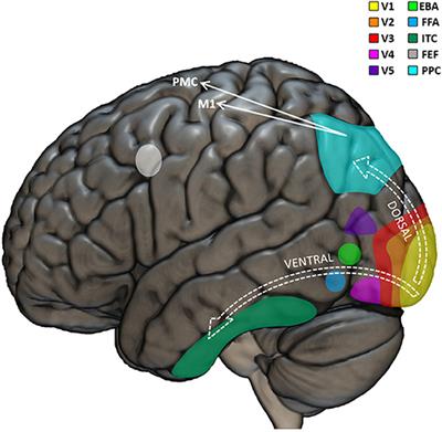
Frontiers | Visual Neuropsychology in Development: Anatomo ...
How Vision Works - BrainHQ from Posit Science 2 weeks ago - Solving the problem of converting light into ideas, of visually understanding features and objects in the world, is a complex task far beyond the abilities of the world’s most powerful computers. Vision requires distilling foreground from background, recognizing objects presented in a wide ...
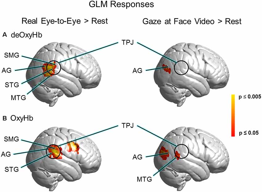
Frontiers | Real-Time Eye-to-Eye Contact Is Associated With ...
Stroke: Rewiring eye-brain connection may restore vision Stroke: Rewiring eye-brain connection may restore vision. Written by Maria Cohut, Ph.D. on March 24, 2019 — Fact checked by Isabel Godfrey. Many people who have a stroke also experience vision ...

3: Diagram of the human brain. Arrows indicate the connection ...
Optic Neuritis and Neuropathy: Symptoms, Causes, Treatments The optic nerve is the connection between the eye and the brain that transmits visual information from the retina. Inflammation of this nerve is called optic neuritis. During optic neuritis inflammation can cause damage to the protective sheath surrounding this nerve and the nerve i

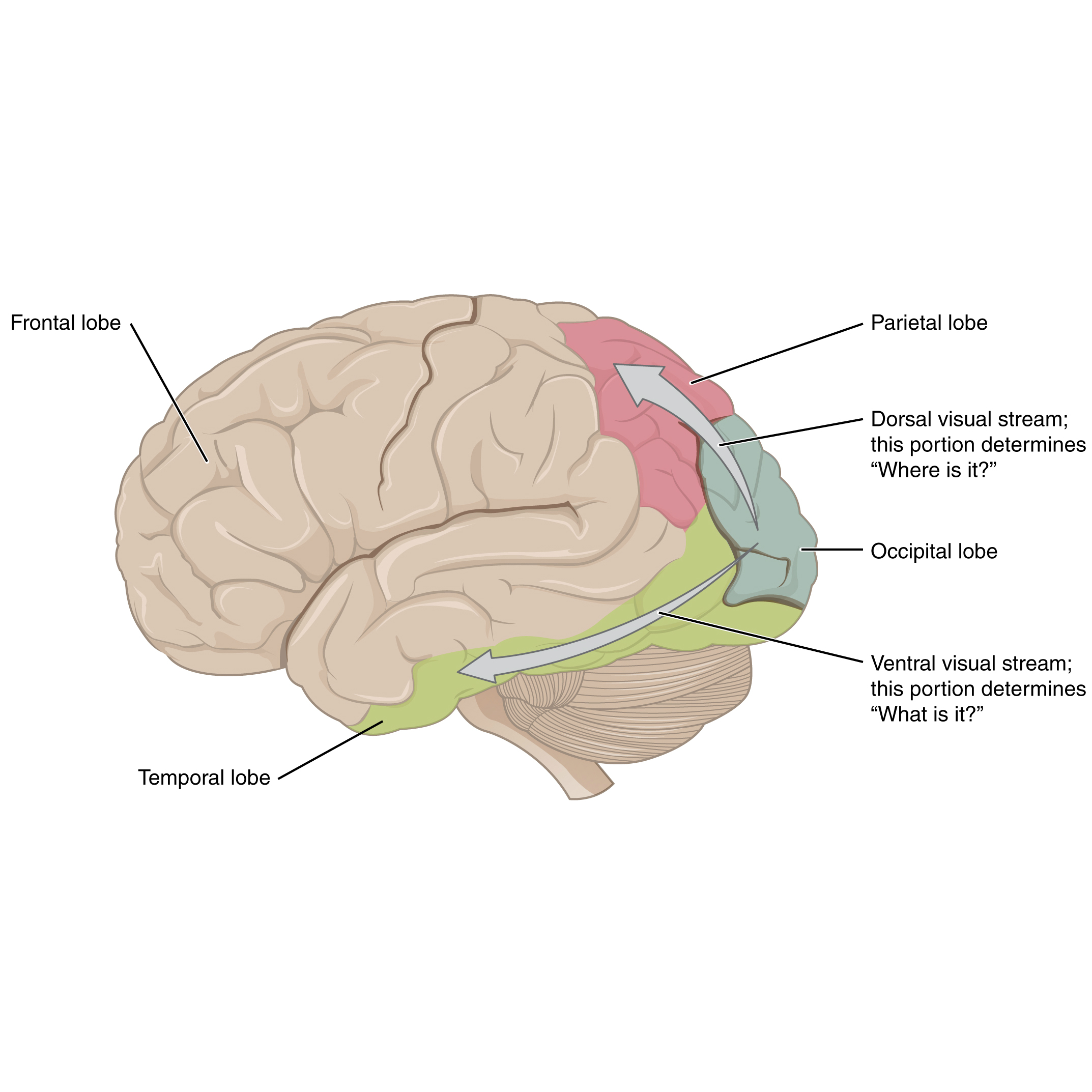
324 Occipital Lobe Illustrations & Clip Art - iStock
Parts Of The Eyelid Diagram - Diagram Sketch Anatomy Of Human Eye Koibana Info. Do You Know The Parts Of The Eye Vocabulary Parts Of The Eye Eye Parts Human Anatomy And Physiology. Click To Close Eye Drawing Diagram Of The Eye Eye Art. Parts Of Eyelid When Applying Eye Makeup Eye Makeup Makeup Grey Smokey Eye.
The Eye
how do the brain and eyes work together - Lisbdnet.com 4 Is the eye directly connected to the brain? 5 What connects the brain to the eye? 6 Do your eyes work together? 7 Do you see with your eyes or brain? 8 How do the eye and brain process visual information? 9 How the brain recognizes what the eye sees? 10 Which part of the brain helps us to see? 11 Are your eyes connected to each other?
THE BRAIN FROM TOP TO BOTTOM
Eye wiring - creation.com Normally, half the nerves of each eye go to each side of the brain, so that each eye is mapped to both the left hemisphere and the right hemisphere (see wiring diagram), but scans on the German girl showed that retinal nerve fibres that should go to the right hemisphere of the brain diverted to the left.

How We See | Introduction to Psychology
THE EYE, THE EAR and the Brain - Sound & Video Contractor PART 1: THE EYE AND THE BRAIN The display of information must meet the needs of the eye. Someof these needs come from the eye itself, and some come from thebrain. THE EYE, AS WE ALL KNOW, IS A LIGHT-sensitive organ of vision thatpermits us to discriminate among minute variations of shape, color,brightness and distance.

The visual pathway from the eye to the brain - Perkins School ...
Eye, Brain, and Vision The fibers corning to the brain from each eye pass uninterrupted through the optic chiasm (from chi, X, the Greek letter whose shape is a cross). There, about half the fibers cross to the side of the brain opposite the eye of origin, and half stay on the same side. From the chiasm the fibers continue to

How Eye Work Medical Illustration, Eye - Brain Diagram, Eye ...
How Does the Eye Work? - Optometrists.org Step 1: Light enters the eye through the cornea When we look at an object, the light that is reflected off of the object enters the eye through the clear front layer of the eye, called the cornea. The cornea bends the light before it passes through a watery substance that fills the area behind the cornea, called the aqueous humor.

Second sight
How Eye Work Medical Illustration, Eye - Brain Diagram ... Cross section through eye and brain showing optic nerve optic chiasma and visual cortex Contains transparencies and gradients How the eye works Cross section of the eye, showing all the major anatomical structures and relationships, including the lens, iris, pupil, cornea and retina. Created in Adobe Illustrator. Contains transparencies. EPS 10.

Visual Pathway Medical Vector Illustration Diagram. Eye And ...
Making connections in the eye: Wiring diagram of retinal ... Citation: Making connections in the eye: Wiring diagram of retinal neurons is first step toward mapping the human brain (2013, August 7) retrieved 11 February 2022 from ...

Visual and biological pathways in the brain: nerve ...
Human Eye Anatomy Stock Photos, Pictures & Royalty-Free ... Parts of the eye, labeled vector illustration diagram How eye work medical illustration, eye - brain diagram, eye... human blue eye extreme macro Eye anatomy. Rod cells and cone cells. Anatomy of human eye hand draw vintage clip art isolated on... eyeball Human eye anatomy vector design Human eye anatomy Eye anatomy isolated on white vector
My Brain, My Eyes, and Cataract Surgery | by Lynn Dorman, Ph ...
Human Brain - Structure, Diagram, Parts Of Human Brain On average, an adult brain weighs between 1.0 kg – 1.5 kg. It is mainly composed of neurons – the fundamental unit of the brain and nervous system. Recent estimates have suggested that the brain contains anywhere between 86 billion to 100 billion neurons. The brain, along with the spinal cord, constitutes the central nervous system. It is ...
Vision | Cochlea
A Very Close Look at the Eye - MIT Technology Review October 22, 2013 - The human brain has 100 billion neurons, connected to each other in networks that allow us to interpret the world around us, plan for the future, and control our actions and movements. MIT neuroscientist Sebastian Seung wants to map those networks, creating a wiring diagram of the brain that ...
Is it possible to actually swap eyes in real life as they do ...
Consumer Updates | FDA Mar 16, 2022 · The .gov means it’s official. Federal government websites often end in .gov or .mil. Before sharing sensitive information, make sure you're on a federal government site.
Making connections in the eye | MIT News | Massachusetts ...
Making connections in the eye | MIT News | Massachusetts ... August 7, 2013 - The human brain has 100 billion neurons, connected to each other in networks that allow us to interpret the world around us, plan for the future, and control our actions and movements. MIT neuroscientist Sebastian Seung wants to map those networks, creating a wiring diagram of the brain that ...

Pin on Βιολογία
The eye is our window to the brain – and there's a lot we ... 18 Mar 2015 — It shows the innermost layer of the eye (the retina), and the nerve carrying visual messages from the retina to the brain (along the optic nerve) ...
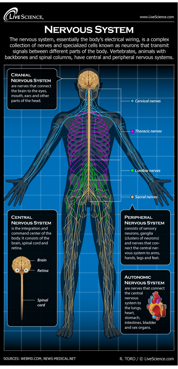
Human Nervous System - Diagram - How It Works | Live Science
optic nerve | anatomy - Encyclopedia Britannica 1 month ago - The optic nerve connects the retina to the visual cortex in the back of the brain. Increased intracranial pressure, tumours, and increased vascular pressure in the eye are possible mechanisms by which the optic nerve can become damaged, impairing vision.Encyclopædia Britannica, Inc.
Brain Anatomy and How the Brain Works | Johns Hopkins Medicine
B. Wiring Diagram of the Visual System - MIT Wiring Diagram of the Visual System Figure 1provides a schematic outline of the primate visual areas and their connectivity. The retinal ganglion cells from the nasal half of each retina send their axons to the contralateral half-brain whereas

How Vision Works - BrainHQ from Posit Science
The Brain and the Eye - How They Work Together - Discovery ... A bundle of more than a million nerve fibers carrying visual messages from the retina to the brain. Your brain actually controls what you see, since it combines images. Also the images focused on the retina are upside down, so the brain turns images right side up. This reversal of the images Is a lot like what a mirror does in a camera.
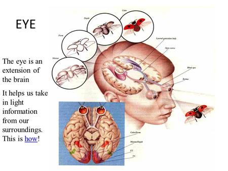
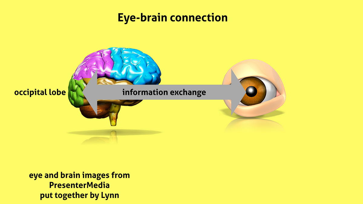
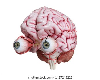
EYE The eye is an extension of the brain. Eye brain proxomity ...
Your Eyes (for Kids) - Nemours KidsHealth The optic nerve serves as a high-speed telephone line connecting the eye to the brain. When you see an image, your eye "telephones" your brain with a report on what you are seeing so the brain can translate that report into "cat," "apple," or "bicycle," or whatever the case may be.

Schematic diagram of the human visual system. The main ...
Eye Diagram Stock Illustrations - 5,953 Eye Diagram Stock ... How eye work medical illustration, eye - brain diagram. Eye structure and connection with brains. Vector EPS10. Eye anatomy diagram,illustration. Eye anatomy diagram,vector illustration. Infographics vector eye design diagram line style timeline. Template.

463 Cranial Nerve Illustrations & Clip Art - iStock
The Eye-Brain Connection The Eye-Brain Connection. Published 18 Jun 2019; Author Michael W. Richardson Source BrainFacts/SfN Light-sensing cells in your retinas transform light and color into electrical signals. But, to turn that information into a complete picture of the world around you, those signals need to be relayed to multiple areas of the brain quickly and ...
The Visual Experience: Reading 2014
Nervous System: Explore the Nerves with Interactive Anatomy ... Nov 02, 2020 · The cranial nerves provide a direct connection to the brain for the special sense organs, muscles of the head, neck, and shoulders, the heart, and the GI tract. Spinal Nerves . Extending from the left and right sides of the spinal cord are 31 pairs of spinal nerves.
Links to Figures and Illustrations Newtonian mechanics Galileo ...
primary visual cortex - THE BRAIN FROM TOP TO BOTTOM Visual acuity is the eye's ability to distinguish two points that are very close to each other. This ability depends on many factors, but especially on the precision of the eye's refraction and the ratio of cones to rods at a given location on the retina · Your eye does exactly the same thing, ...
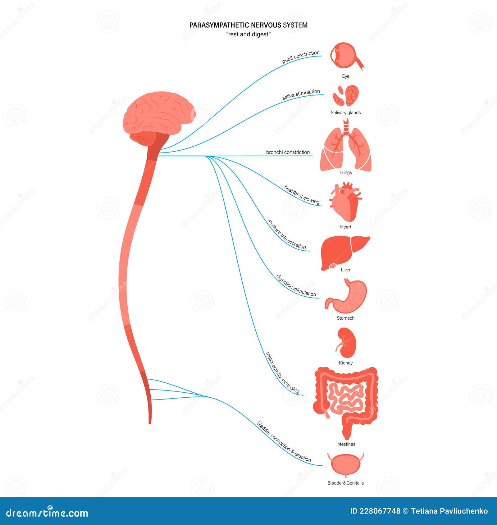
Parasympathetic Nervous System Stock Vector - Illustration of ...
Vision | Introduction to Psychology Rods and cones are connected (via several interneurons) to retinal ganglion cells. Axons from the retinal ganglion cells converge and exit through the back of the eye to form the optic nerve. The optic nerve carries visual information from the retina to the brain.
File:The Process of Bionic Eyes for the Visually Impaired.svg ...
How Eye Work Medical Illustration Eye Brain Diagram Eye ... Description How eye work medical illustration, eye - brain diagram, eye structure and connection with brains. Vector EPS10 1 credit Essentials collection for this image $4 with a 1-month subscription (10 Essentials images for $40) Continue with purchase View plans and pricing Includes our standard license. Add an extended license.
Eye and extraocular muscles (illustrations) | Image ...
The visual pathway from the eye to the brain - Perkins ... Graphic of visual pathway looking down onto the brain: eyes, optic nerves, optic. Here are the key points along the route: Retina: This is your eye's on-ramp.

Autonomic nervous system stock vector. Illustration of lungs ...
Anatomy of the Eye | Kellogg Eye Center | Michigan Medicine The, clear, gelatinous substance filling the central cavity of the eye. How the Eye Works. The five senses include sight, sound, taste, hearing and touch. Sight, like the other senses is closely related to other parts of our anatomy. The eye is connected to the brain and dependent upon the brain to interpret what we see.
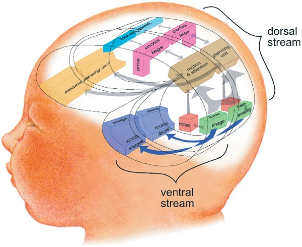
Cognitive vision, its disorders and differential diagnosis in ...
The Optic Nerve And Its Visual Link To The Brain ... The Optic Nerve And Its Visual Link To The Brain The optic nerve, a cable-like grouping of nerve fibers, connects and transmits visual information from the eye to the brain. The optic nerve is mainly composed of retinal ganglion cell (RGC) axons.
Breakthrough Discovery Leads to Invention of neurolenses® –
Visualizing the Connections Between Eye and Brain ... In this study published May 31 in the journal Cell, Mark Andermann, PhD, Chinfei Chen, MD, PhD, and colleagues developed a means of tracking the activity of the far-reaching ends of retinal neurons (called boutons) as they deliver visual information to the thalamus, a brain region involved in image processing.
Vision: It all Starts with Light
Wiring diagram of retinal neurons is first step toward mapping ... 1 month ago - Massachusetts Institute of Technology. (2013, August 7). Making connections in the eye: Wiring diagram of retinal neurons is first step toward mapping the human brain. ScienceDaily. Retrieved March 8, 2022 from
Cornea Function, Definition & Anatomy | Body Maps
Human eye - Wikipedia The approximate field of view of an individual human eye (measured from the fixation point, i.e., the point at which one's gaze is directed) varies by facial anatomy, but is typically 30° superior (up, limited by the brow), 45° nasal (limited by the nose), 70° inferior (down), and 100° temporal (towards the temple).

Human Visual System - an overview | ScienceDirect Topics
A Wiring Diagram of the Brain - MIT Technology Review Only one organism's wiring diagram currently exists: that of the microscopic worm C. elegans. Despite containing a mere 302 neurons, the C. elegans mapping effort took more than a decade to...

Visual system - Wikipedia
Our Sense of Sight: Part 1. Eye Anatomy and Function Eye Anatomy and Function · FEATURING: A "CLASS EXPERIMENT" PLUS: "TRY YOUR OWN EXPERIMENT"
Eye and brain Images, Stock Photos & Vectors | Shutterstock
0 Response to "37 eye to brain connection diagram"
Post a Comment