39 on the diagram below identify alveolar epithelium
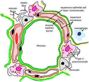
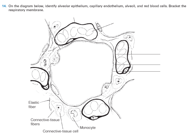
essayhelpp.com › biology-lBIOLOGY L - Essay Help Mar 07, 2022 · The diagram below represents a single atom of hydrogen; From genetic engineering to gene editing to nanotechnology, a number of biotechnology approaches are currently being used or being developed for use to improve the quality and quantity of our food sup… OBSERVATIONS AND DATA 1. What is the variable in this experiment? 2. ASSIGNMENT 2. KNOWLEDGE LEARNED.docx - 1. 2. 3. 4. 5. 6. 7 ... On the diagram below, identify alveolar epithelium, capillary endothelium, alveoli, and red blood cells. Bracket the respiratory membrane 1. Alveoli (gas-filled air spaces) 2. Red Blood cell in capillary 3. Type II alveolar cell (surfactant secreting) 4. Type I alveolar cell 5. Respiratory membrane 6. Endothelial cell nucleus 7. Macrophage 8.
ecampusontario.pressbooks.pub › medicalterminologyRespiratory System – Building a Medical Terminology Foundation The epithelium contains specialized epithelial cells that produce mucus to trap debris. The cilia of the respiratory epithelium help to remove mucus and debris with a constant beating motion, sweeping materials towards the throat to be swallowed. This moist epithelium functions to warm and humidify incoming air.

On the diagram below identify alveolar epithelium
4.2 Epithelial Tissue - Anatomy & Physiology Figure - 4.2.3 Goblet Cell: (a) In the lining of the small intestine, columnar epithelium cells are interspersed with goblet cells. (b) The arrows in this micrograph point to the mucous-secreting goblet cells (LM × 1600). (Micrograph provided by the Regents of University of Michigan Medical School © 2012) External Website The micromechanics of lung alveoli: structure and function ... The alveolar epithelium (thin type I cell extension marked by arrowheads) is covered with a lining layer containing intra-alveolar surfactant (Surf). Alv alveolar lumen. Scale bar 1 µm. Inset shows tubular myelin, a surface-active intra-alveolar surfactant subtype, at higher magnification. Scale bar 0.5 µm Alveoli Flashcards - Quizlet - The epithelial lining of alveoli consists mainly of type I alveolar cells (also known as type I pneumocytes). - These are large, flat, squamous cells with few organelles and thin cytoplasm. - They cover about 93% of alveolar surface area. - Their primary purpose is air-blood gas exchange. - The junctions between these cells are narrow (1nm).
On the diagram below identify alveolar epithelium. › quiz-school › topic35 Histology Quizzes Online, Trivia, Questions ... - ProProfs Mar 22, 2022 · A comprehensive database of more than 35 histology quizzes online, test your knowledge with histology quiz questions. Our online histology trivia quizzes can be adapted to suit your requirements for taking some of the top histology quizzes. Answered: Identify the structures on the diagram.… | bartleby On the diagram below, identify alveolar epithelium, capillary endothelium, alveoli, and red bloo... A: Lungs provide the respiratory surface for the exchange of gases. Alveoli are very small sacs present... quizlet.com › 143609117 › a-p-ii-chapter-22-labA & P II Chapter 22 Lab Flashcards - Quizlet Recall that the air in the lungs can be divided into respiratory volumes and capacities. Drag the labels to identify the average respiratory volumes and capacities for a healthy, adult male on the spirometry tracing shown below. Solved iagram below, identify alveolar epithelium ... Expert Answer 100% (3 ratings) Ans. 1- Alveolar epithelium 2- Capil … View the full answer Transcribed image text: iagram below, identify alveolar epithelium, capillary endothelium, alveoli, and red blood cells. Bracket the brane respira Elastic fiber Connective tissue fibers Monocyte Previous question Next question
Human Structure Virtual Microscopy - IU Note that the oral epithelium is thicker. Locate the small salivary glands in the submucosa of the lip and the skeletal muscle. Examine the diagram below and this section of an adult tooth, which is from a decalcified tooth still embedded in the bone of the jaw. The enamel, a calcified material similar to bone, is completely dissolved away. The Respiratory System - Chapter 23 Flashcards | Quizlet alveolar epithelium The respiratory membrane is a composite structure consisting of three layers: (1) the endothelial cells lining an adjacent capillary (capillary endothelium) (2) the fused basal laminae that lie between the alveolar and endothelial cells(basement membrane - fused basal lamiae) Respiratory System | histology B. Alveolar ducts The walls of alveolar ducts View Image are lined by alveoli and alveolar sacs (clusters of alveoli). C. Alveolus The walls of these structures are covered on both sides by squamous epithelium (too thin to see) of Type I cells lining adjacent alveolar lumens. Within the walls is an extensive capillary network. Epithelial Tissue: Structure with Diagram, Function, Types ... Epithelial cells form membranes. The epithelial membrane consists of a layer of epithelial tissue and has underlying connective tissue. There are two types of epithelial membranes, mucous membrane and serous membrane. Mucous membrane: It is also known as mucosa. There are goblet cells present, which secrete mucus.
Respiratoryanatomy - description - BIO-120-2323 ... alveolar sac. middle lobe. terminal bronchiole. respiratory bronchioles. inferior lobe alveolar duct. alveolar duct alveoli ##### 13. On the diagram below identify alveolar epithelium, capillary endothelium, alveoli, and red blood cells. Bracket the ##### respiratory membrane. Demonstrating Lung Inflation in a Sheep Pluck ##### 14. 22.1 Organs and Structures of the Respiratory System ... A bronchial tree (or respiratory tree) is the collective term used for these multiple-branched bronchi. The main function of the bronchi, like other conducting zone structures, is to provide a passageway for air to move into and out of each lung. In addition, the mucous membrane traps debris and pathogens. Structure and function of the alveolus - Deranged Physiology Structure and shape of the alveoli. Alveoli are the basic unit of the gas exchange surface. In summary, the following things can be said about the alveolar shape and structure: Largely polyhedral shape. Open at one end, like a cup. Walls of the alveoli are composed of the pulmonary capillary sheet. PDF A-level BIOLOGY 7402/1 The answer space for the diagram is on the opposite page. [2 marks] 13 *13* Space for diagram: [Turn over] 10 * 14 * 14 0 2 To study lipid digestion, a scientist placed a tube into the gut of a healthy 20 - year - old man. The end of the tube passed through the stomach but did not reach as far as the ileum. The scientist fed the man a meal

Respiratory A&P
PDF Practice Quiz Tissues Identify the tissue type and a location where it is found. Simple Squamous Epithelium •Kidney glomeruli • Air sacs of lungs • Serosae •Lining of heart, blood vessels and lymphatic vessels Identify the structure indicated. Compact Bone (Osseous Tissue) Interstitial Lamellae Identify the structure indicated. Simple Squamous Epithelium: Nucleus

Uncategorized Archives - Page 2 of 8 - Respiratory Learning
Epithelial tissue: definition, functions & examples | Kenhub Epithelium is one of only 4 types of human body tissues.Like all types, it is formed by cells within an extracellular matrix (ECM). The cells in this tissue are tightly packed within a thin ECM. Forming sheets that cover the internal and external body surfaces (surface epithelium) and secreting organs (glandular epithelium). Functions of epithelial tissue are secretion, protection, absorption ...

COVID-19 and Respiratory System Disorders | Arteriosclerosis ...
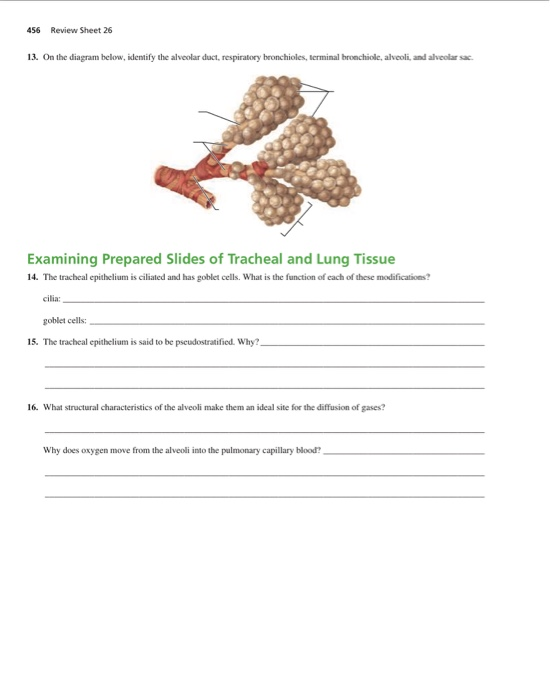
Solved 13. On the diagram below, identify the alveolar ... On the diagram below, identify the alveolar duct, respiratory bronchioles, terminal bronchiole, alveoli, and alveolar saci Examining Prepared Slides of Tracheal and Lung Tissue 14. The tracheal epithelium is ciliated and has goblet cells. What is the function of each of these modifications? cilia: goblet cells: 15.
Host metabolism dysregulation and cell tropism identification ...
Histology Slides 1 Every surface facing the alveolar air spaces is lined by simple squamous epithelium of the alveolar walls. Slide 17 Detail of the septa between alveolar spaces. The small spaces within the septa are empty capillary lumens. Nuclei belong mainly to endothelial cells and alveolar epithelium; it would take EM to identify which is which with certainty.
Anatomy of the Respiratory System
Alveoli Definition, Location, Anatomy, Function, Diagrams Alveoli are the small balloon-like sacks of 200-500μm diameter [1], making up a vital part of the respiratory zone of the human lungs. Each alveolus (singular) plays an important role in letting oxygen and carbon dioxide move into and from the bloodstream during inhalation and exhalation [2, 3]. Where are the alveoli found
Cells | Free Full-Text | Extracellular Lipids in the Lung and ...
Alveoli: Anatomy, function and clinical points - Kenhub The pulmonary alveolus is a sac roughly 0.2 to 0.5 mm in diameter. These alveoli are located at the ends of air passageways in the lungs. Sometimes, people compare alveoli structures to the appearance of a raspberry or a "bunch of grapes."

Identification of alveolar, adventitial, and peribronchial ...
PDF AS 17 TISSUES X Y Z - WordPress.com (b) The diagram below shows a longitudinal section through a striated muscle fibril as seen under the electron microscope. Same fibril under light microscope (i) Draw in the details of the fibril (between the parallel lines) to show its appearance under the light microscope. [2] (ii) Identify parts 1 to 6 shown in the diagram.

Alveolar epithelial cells during alveolarization. During the ...
Exercise23.pdf - Exercise 23 Review Sheet Anatomy of the ... Describe the appearance of a pseudostratified epithelium. It is thick and appears to be composed of more than one layer. 1. On the diagram below, identify alveolar duct, respiratory bronchioles, terminal bronchiole, alveoli, and alveolar sac. 2. 3. By what process does oxygen move from the alveolar sac into the blood in a pulmonary capillary?

Transcriptional Mechanisms Regulating Alveolar Epithelial ...
› biology › human-respiratory-systemHuman Respiratory System - Diagram, Features, Parts and Functions Human Respiratory System Diagram. If you carefully observe the respiratory system diagram, you will be able to see the various organs involved in its functioning. (Image will be Uploaded soon) Features of the Human Respiratory System. The structure of the lungs is created in such a way that it helps the exchange of gasses.

History of lung organoids. The important events in the ...
en.wikipedia.org › wiki › PeriodontologyPeriodontology - Wikipedia The junctional epithelium provides a specialised protective barrier to microorganisms residing around the gingival sulcus. Collagen fibres bind the attached gingiva tightly to the underlying periodontium including the cementum and alveolar bone and vary in length and width, [4] depending on the location in the oral cavity and on the individual.

Organs and Structures of the Respiratory System | Anatomy and ...
PDF (1) - WordPress.com alveolar epithelium to become thicker. People with miner's lung have a lower concentration of oxygen in their blood than healthy people. (a)€€€€ (i)€€€€€ Describe the path by which oxygen goes from an alveolus to the blood.
Autophagic flux blockage in alveolar epithelial cells is ...
study.com › academy › lessonAdipose Tissue and Loose Connective Tissue ... - Study.com Aug 24, 2021 · The amount of the three fiber types varies in this tissue depending on where it is located and the job it performs. Therefore, the appearance of areolar connective tissue varies depending on where ...

Uncategorized Archives - Page 2 of 8 - Respiratory Learning
Answered: 14. On the diagram below, identify… | bartleby 14. On the diagram below, identify alveolar epithelium, capillary endothelium, alveoli, and red blood cells. Bracket the respiratory membrane. Elastic - fiber Connective-tissue fibers Monocyte Connective-tissue cell Question Transcribed Image Text: 14.

Identification of alveolar epithelial type II cells and ...
Alveolar Epithelium - an overview | ScienceDirect Topics Diagram of alveolus and ion channels, pumps, and pores in the alveolar epithelium. The alveolus is composed of alveolar epithelial type 1 cells, alveolar epithelial type 2 cells and capillary cells. Type 1 cells are large, squamous epithelial cells that cover ~95% of the alveolar surface area.

Alveoli form directly by budding led by a single epithelial ...
A revised model of alveolar regeneration We identify ... A revised model of alveolar regeneration We identify convergence of alveolar and airway stem cells on an injury-induced transitional cell state characterized by a unique transcriptional signature,...

Pulmonary alveolus - Wikipedia
S9) The Respiratory System Flashcards by ... - Brainscape There are epithelial changes in the respiratory system. What sort of epithelium can be found in the nasal cavity, pharynx, larynx, trachea, primary bronchi and secondary bronchi? ... Identify some key anatomical features on the larynx diagram below: 19 ... - Alveolar walls are damaged, bronchioles collapse during exhalation, so lungs cannot ...
Alveoli: Anatomy, function and clinical points | Kenhub
Epithelial Tissue | Anatomy and Physiology I In tubular glands, the ducts can be straight or coiled, whereas tubes that form pockets are alveolar (acinar), such as the exocrine portion of the pancreas. Combinations of tubes and pockets are known as tubuloalveolar (tubuloacinar) compound glands. In a branched gland, a duct is connected to more than one secretory group of cells. Figure 4.

Pneumocystis carinii Cell Wall β-Glucan Induces Release of ...
Respiratory: The Histology Guide This diagram shows a diagram of an alveolar sac, showing how the organisation of the alveoli, and the network of blood capillaries that surround the alveoli (in red). The epithelium of the alveoli, contains two main types of cells:
In vitro modelling of alveolar repair at the air-liquid ...
Alveoli Flashcards - Quizlet - The epithelial lining of alveoli consists mainly of type I alveolar cells (also known as type I pneumocytes). - These are large, flat, squamous cells with few organelles and thin cytoplasm. - They cover about 93% of alveolar surface area. - Their primary purpose is air-blood gas exchange. - The junctions between these cells are narrow (1nm).

STAT3–BDNF–TrkB signalling promotes alveolar epithelial ...
The micromechanics of lung alveoli: structure and function ... The alveolar epithelium (thin type I cell extension marked by arrowheads) is covered with a lining layer containing intra-alveolar surfactant (Surf). Alv alveolar lumen. Scale bar 1 µm. Inset shows tubular myelin, a surface-active intra-alveolar surfactant subtype, at higher magnification. Scale bar 0.5 µm

COVID-19 and Respiratory System Disorders | Arteriosclerosis ...
4.2 Epithelial Tissue - Anatomy & Physiology Figure - 4.2.3 Goblet Cell: (a) In the lining of the small intestine, columnar epithelium cells are interspersed with goblet cells. (b) The arrows in this micrograph point to the mucous-secreting goblet cells (LM × 1600). (Micrograph provided by the Regents of University of Michigan Medical School © 2012) External Website

Patrolling Alveolar Macrophages Conceal Bacteria from the ...

Comprehensive epigenomic profiling of human alveolar ...

Biology Notes for A level: #51 Summary of Gas exchange

Alveolar Epithelium - an overview | ScienceDirect Topics

1: Histology of the alveolar capillary barrier. The alveolar ...

Biology Notes for A level: #82 Question 5

Alveoli form directly by budding led by a single epithelial ...

Crosstalk between alveolar macrophages and alveolar ...
Untitled

Acquisition of cellular properties during alveolar formation ...

Alveolar wars: The rise of in vitro models to understand ...
Solved 456 Review Sheet 26 13. On the diagram below, | Chegg.com
Regulation of immune responses by the airway epithelial cell ...
Untitled
Respiratory: The Histology Guide

Alveolar epithelial type I cell morphology. (A) Scanning ...
A cross-talk between epithelium and endothelium mediates ...
Answered: 14. On the diagram below, identify… | bartleby
0 Response to "39 on the diagram below identify alveolar epithelium"
Post a Comment