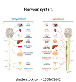
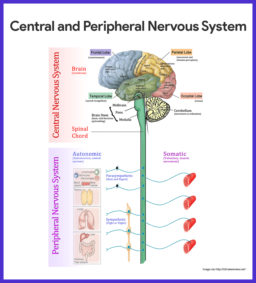
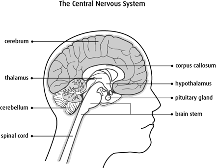
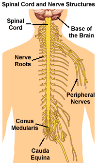
36 brain and spinal cord diagram
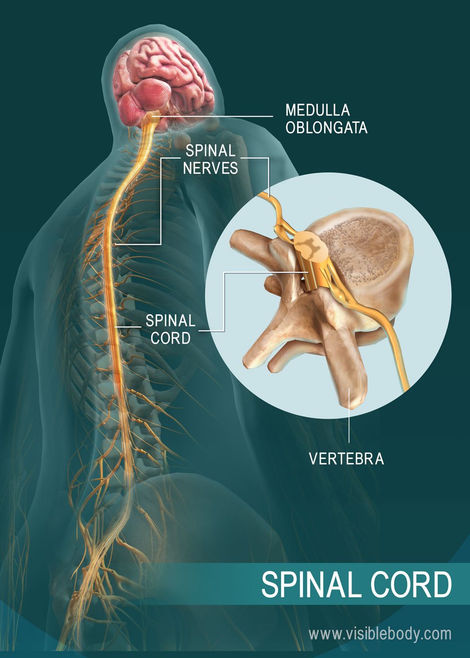
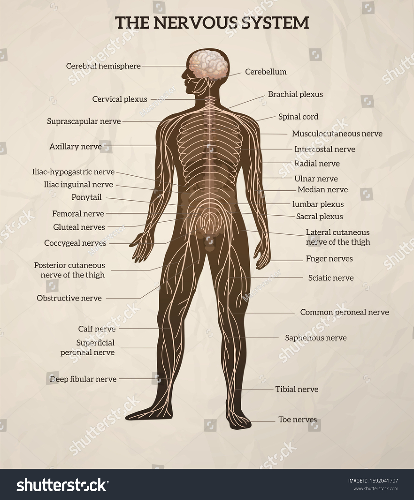
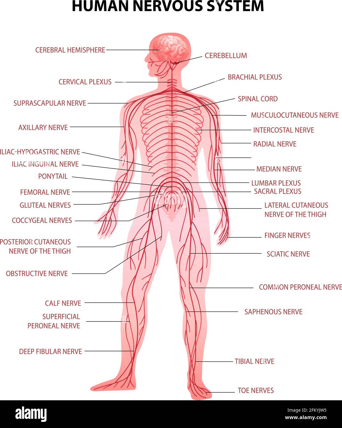
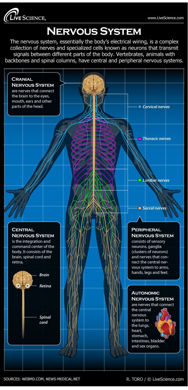
A Neurosurgeon's Overview of the Anatomy of the Spine and ... The spinal cord is an extension of the central nervous system (CNS), which consists of the brain and spinal cord. The spinal cord begins at the bottom of the brain stem (at the area called the medulla oblongata) and ends in the lower back, as it tapers to form a cone called the conus medullaris.. Anatomically, the spinal cord runs from the top of the highest neck bone (the C1 vertebra) to ... Diagram of the brain and spinal cord. Diagram of the brain ... diagram of the brain and spinal cord illustrating how the eye's inner and outer blood-retinal barriers (brbs) fit into the overall scheme of blood-neural barriers (bnbs) barriers between the blood...
Spinal Cord Diagram with Detailed Illustrations and Clear ... Spinal Cord Diagram Spinal Cord Diagram The spinal cord is one of the most important structures in the human body. In fact, it is the most important structure for any vertebrates. Anatomically, the spinal cord is made up is made up of nervous tissue and is integrated into the spinal column of the backbone. Main Article:
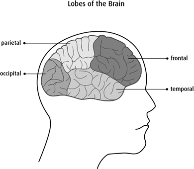
Brain and spinal cord diagram
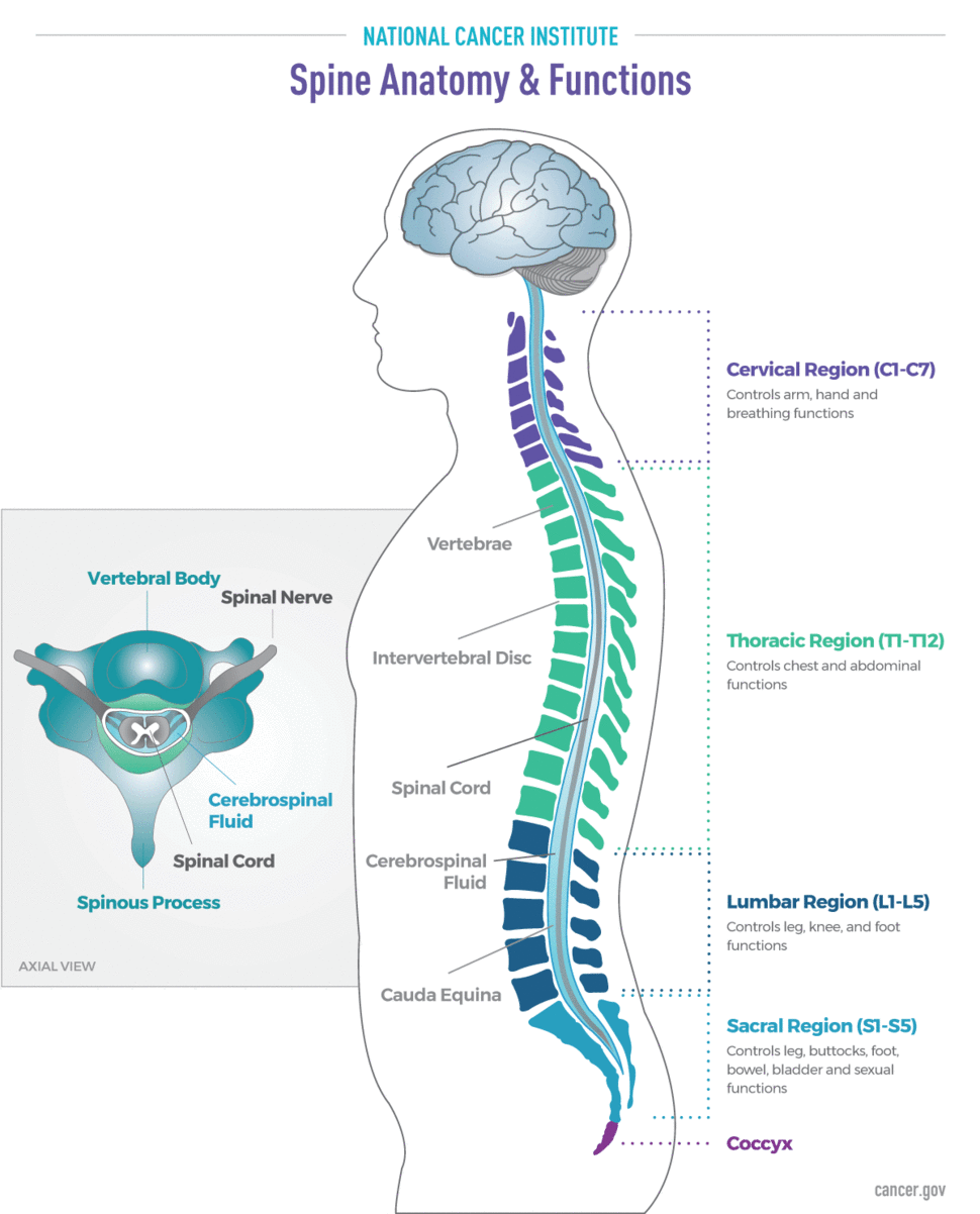
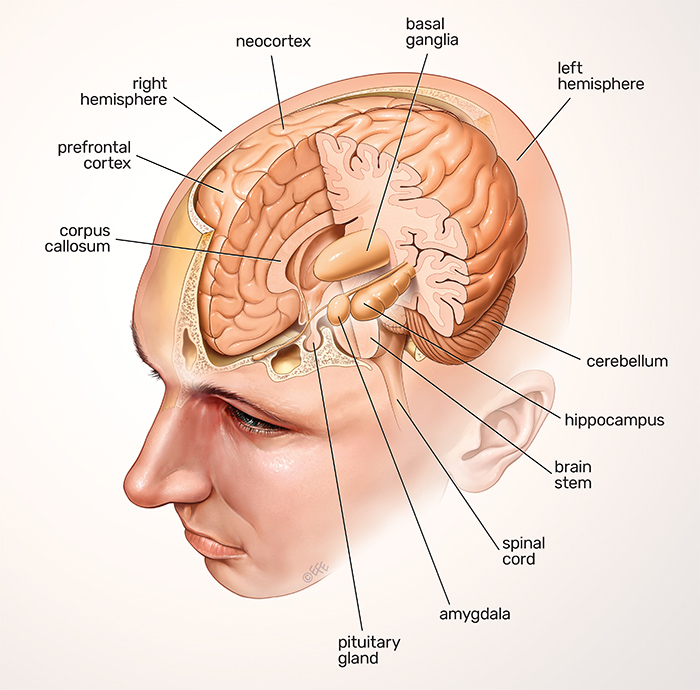
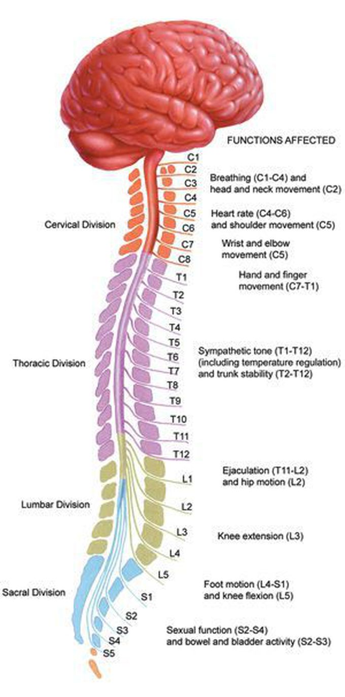
Spinal Cord - Anatomy, Structure, Function, & Diagram Spinal Cord Anatomy In adults, the spinal cord is usually 40cm long and 2cm wide. It forms a vital link between the brain and the body. The spinal cord is divided into five different parts. Sacral cord Lumbar cord Thoracic cord Cervical cord Coccygeal Several spinal nerves emerge out of each segment of the spinal cord. Spinal Cord Quiz: Cross-Sectional Anatomy - GetBodySmart Spinal Cord Quiz: Cross-Sectional Anatomy. Learn anatomy faster and remember everything you learn. Start Now. Spinal Cord - Cross-Sectional Anatomy. Start Quiz ... Parts of the Brain Quiz. Test your knowledge with the parts of the brain and their functions in a fun and interactive way. Click and start your quiz immediately! 14.3 The Brain and Spinal Cord - Anatomy & Physiology The spinal cord is a single structure, whereas the adult brain is described in terms of four major regions: the cerebrum, the diencephalon, the brain stem, and the cerebellum. A person's conscious experiences are based on neural activity in the brain. The regulation of homeostasis is governed by a specialized region in the brain.
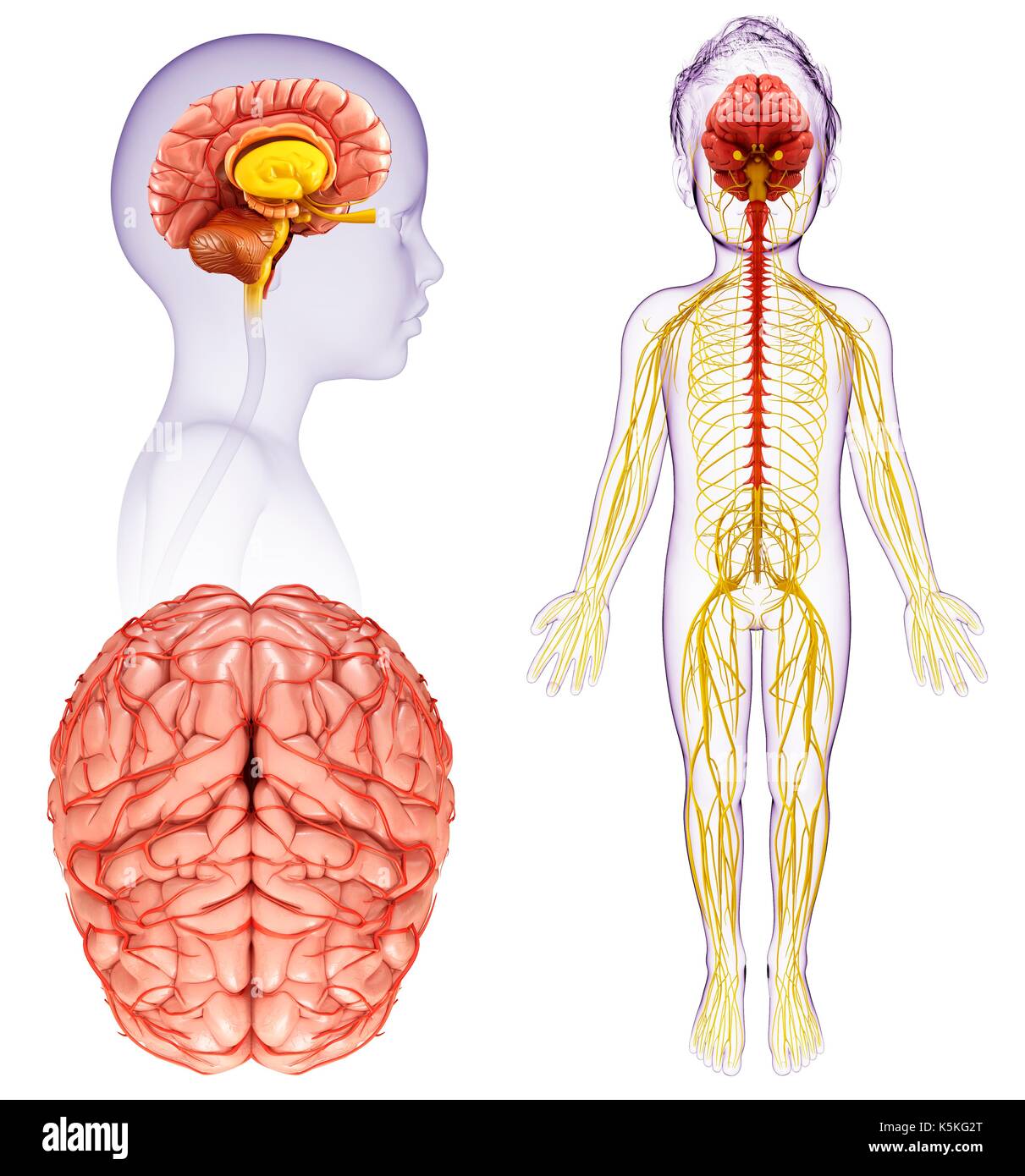
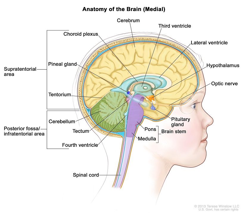
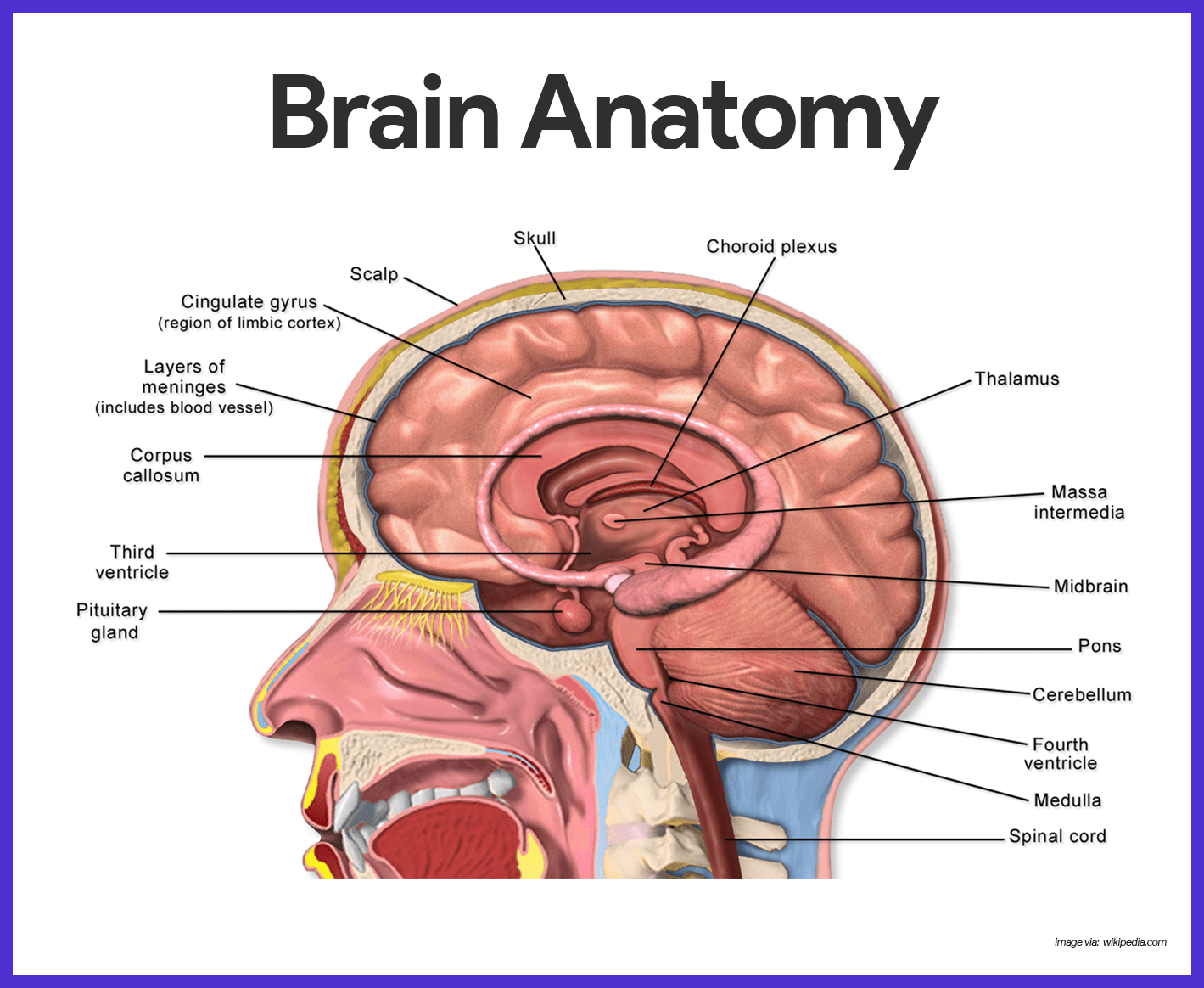
Brain and spinal cord diagram. Ventricles of the Brain Explained With a Diagram - Bodytomy Ventricles are hollow cavities of the brain, that contain the cerebrospinal fluid (CSF), which circulates within the brain and spinal cord. There are all together four ventricles in the human brain, that constitute the ventricular system, along with the cerebral aqueduct. They are known as, lateral ventricles, third ventricle, and fourth ventricle. BRAIN AND SPINAL CORD Diagram | Quizlet Start studying BRAIN AND SPINAL CORD. Learn vocabulary, terms, and more with flashcards, games, and other study tools. Spinal Cord Diagram | Spinal Cord Facts | DK Find Out The spinal cord is the major link between the brain and the rest of the body. Although it is only as wide as a little finger, it contains more than 20 million nerve fibers. From it extend 31 pairs of spinal nerves to the chest, arms, lower body, and legs. An adult's spinal cord is about 17in (43cm)long. Human Body›Brain and nerves›Spinal cord› Diagram of the Brain and its Functions - Bodytomy The extension of the central nervous system is through the medulla oblongata, which continues as the spinal cord, by exiting the skull through the foramen magnum and continuing in the spinal column. This is where the spinal cord gives out nerves which form the peripheral nervous system.
The Nervous System: Brain and Spinal Cord Diagram | Quizlet The Nervous System: Brain and Spinal Cord STUDY PLAY central nervous system (CNS) consists of the brain and spinal cord; body's integration and interpretation center the CNS is composed mainly of interneurons peripheral nervous system (PNS) conssits of all parts of the nervous system outside the brain and spinal cord the PNS is composed mainly of The Human Brain - Diagram, Parts and Function of Human Brain The spinal cord is active when the brain is busy. It is also called the 2 nd brain of the human body. It begins in continuation with the medulla oblongata and extends; The spinal cord is enclosed in a bony cage called vertebral; A total of thirty-one pairs of spinal nerves arise from the spinal; Functions of Spinal Cord Human Spinal Cord: Structure and Effects (With Diagram) These fibers reach posterior funiculus of spinal cord and ascend up on same side of spinal cord. In the brainstem, at the level of medulla oblongata, these fibers synapse in two different nuclei, namely gracile and cuneate nuclei. First order neurons are posterior root ganglion cells. Spinal cord: Anatomy, structure, tracts and function | Kenhub The spinal cord is made of gray and white matter just like other parts of the CNS. It shows four surfaces: anterior, posterior, and two lateral. They feature fissures (anterior) and sulci (anterolateral, posterolateral, and posterior). The gray matter is the butterfly-shaped central part of the spinal cord and is comprised of neuronal cell bodies.
Brain - Human Brain Diagrams and Detailed Information Connecting the brain to the spinal cord, the brainstem is the most inferior portion of our brain. Many of the most basic survival functions of the brain are controlled by the brainstem. The brainstem is made of three regions: the medulla oblongata, the pons, and the midbrain. Brain, spinal cord and peripheral nervous system anatomy ... The brain is found in the cranial cavity, while the spinal cord is found in the vertebral column. Both are protected by three layers of meninges (dura, arachnoid, and pia mater). The brain generates commands for target tissues and the spinal cord acts as a conduit, connecting the brain to peripheral tissues via the PNS. Spinal Cord: Function, Anatomy and Structure The spinal cord is a long, tube-like band of tissue. It connects your brain to your lower back. Your spinal cord carries nerve signals from your brain to your body and vice versa. These nerve signals help you feel sensations and move your body. Any damage to your spinal cord can affect your movement or function. Appointments 866.588.2264 Spinal Cord | Interactive Anatomy Guide The spinal cord is a major component of the central nervous system (CNS) that forms the vital link between the brain and most of the body. Its 31 spinal segments connect the CNS to the organs and tissues of the neck, torso, and limbs. The spinal cord also performs important processing functions to maintain balance and respond quickly to stimuli.
14.3 The Brain and Spinal Cord - Anatomy & Physiology The spinal cord is a single structure, whereas the adult brain is described in terms of four major regions: the cerebrum, the diencephalon, the brain stem, and the cerebellum. A person's conscious experiences are based on neural activity in the brain. The regulation of homeostasis is governed by a specialized region in the brain.
Spinal Cord Quiz: Cross-Sectional Anatomy - GetBodySmart Spinal Cord Quiz: Cross-Sectional Anatomy. Learn anatomy faster and remember everything you learn. Start Now. Spinal Cord - Cross-Sectional Anatomy. Start Quiz ... Parts of the Brain Quiz. Test your knowledge with the parts of the brain and their functions in a fun and interactive way. Click and start your quiz immediately!
Spinal Cord - Anatomy, Structure, Function, & Diagram Spinal Cord Anatomy In adults, the spinal cord is usually 40cm long and 2cm wide. It forms a vital link between the brain and the body. The spinal cord is divided into five different parts. Sacral cord Lumbar cord Thoracic cord Cervical cord Coccygeal Several spinal nerves emerge out of each segment of the spinal cord.


/brain_spinal_cord-57fe96b15f9b5805c26d5072.jpg)



:background_color(FFFFFF):format(jpeg)/images/library/13921/Central_nervous_system.png)

(246).jpg)








0 Response to "36 brain and spinal cord diagram"
Post a Comment