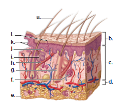
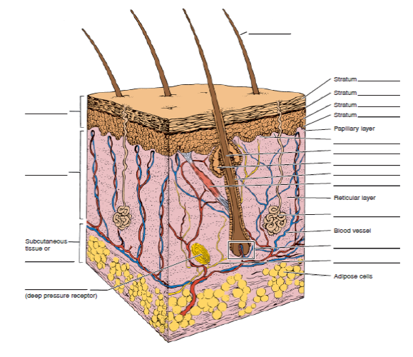
34 skin structure diagram to label
The skin is the largest organ of the body, covering an area of approximately 2 m2. The skin is composed of the cutis (including the dermis and epidermis ), subcutaneous tissue , and skin appendages. The epidermis , which is derived from ectoderm , is the outermost layer of the skin and is mainly composed of keratinocytes . The dermis
The skin was previously viewed as a body part that protects us from the elements. Today, new knowledge informs us that the layers of the skin are actually very complex and have many important functions—from giving us goosebumps and cooling us down in the sauna to letting our brain know that our hand is on a burner.
Skin forms a physical barrier between the inside of an organism and its external environment Structure of the Skin The skin consists of three major layers; the epidermis, the dermis, and the hypodermis (AKA the subcutaneous layer ). The epidermis is the outermost layer of the skin.

Skin structure diagram to label
1. The vulva. It's a common misconception that the visible outer parts of the female anatomy is called the vagina. The technical name is actually the vulva. Yours has the job of protecting your ...
34,524 skin layers stock photos, vectors, and illustrations are available royalty-free. See skin layers stock video clips. of 346. plastic bottle layers of dermis acne illustration acne skin structure structure of the skin anatomy skin an layersskin diagrams epidermis tissue skin structure skin section. Try these curated collections.
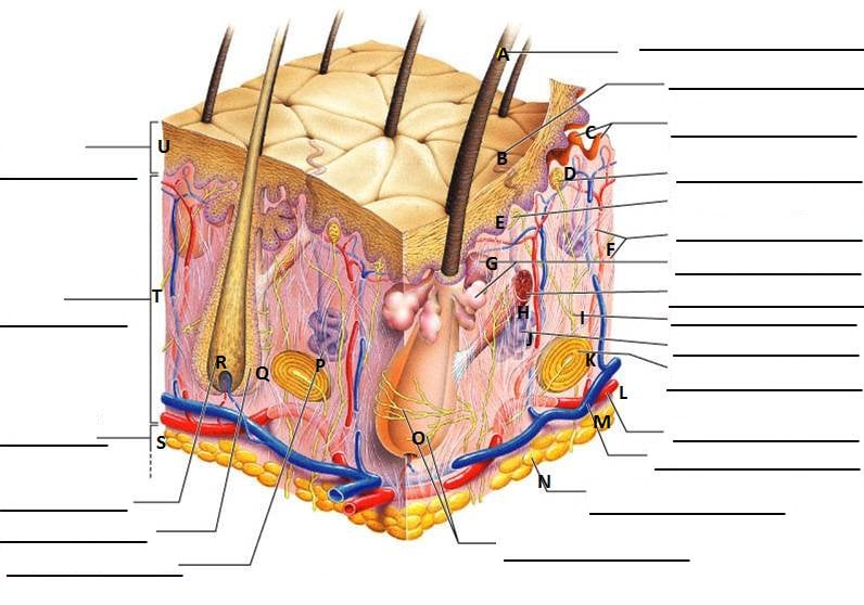
Label the diagram with the letters below according to the structure/area they describe. You may label with a line or put the label directly onto the area ...3 pages
Skin structure diagram to label.
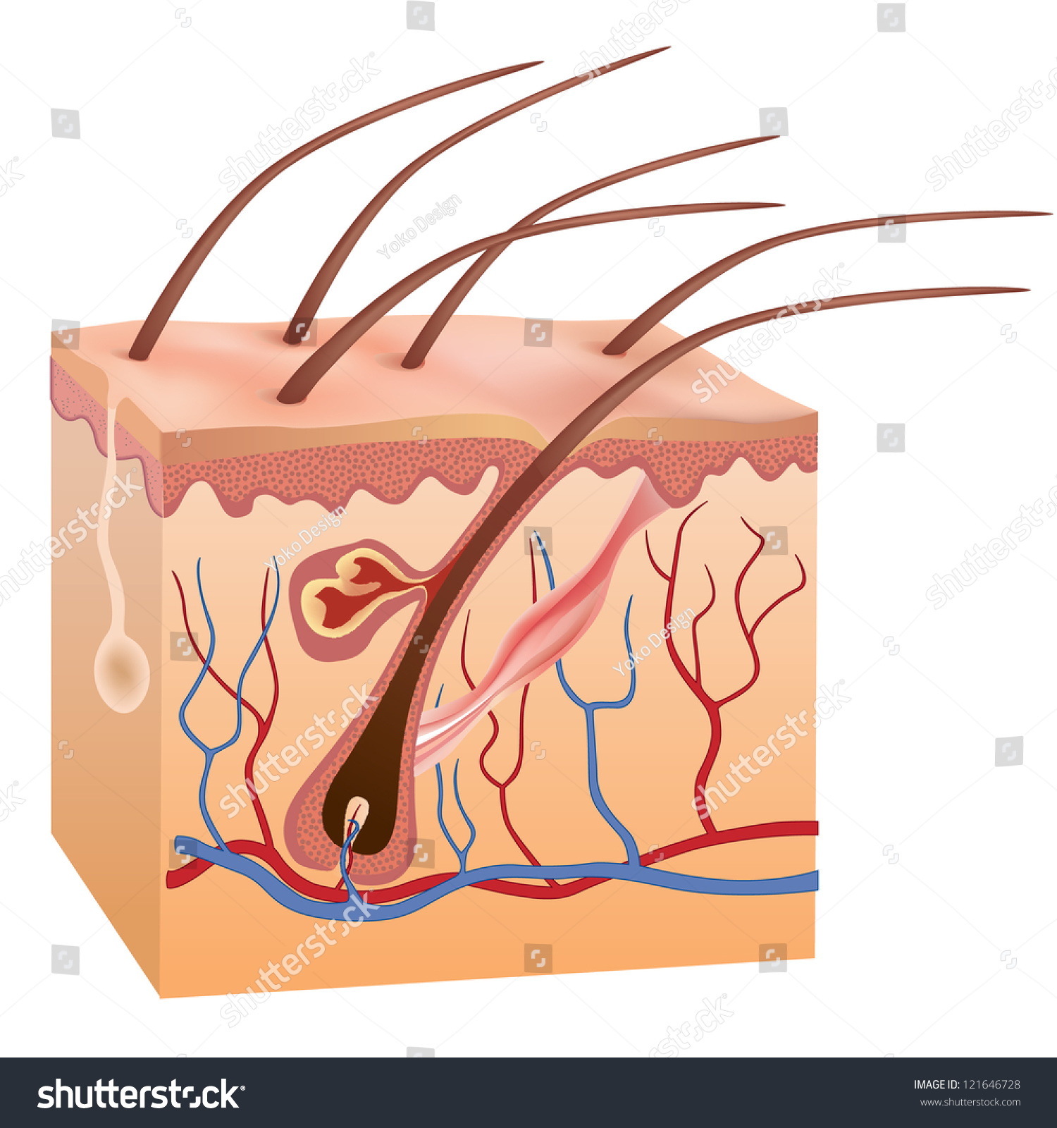
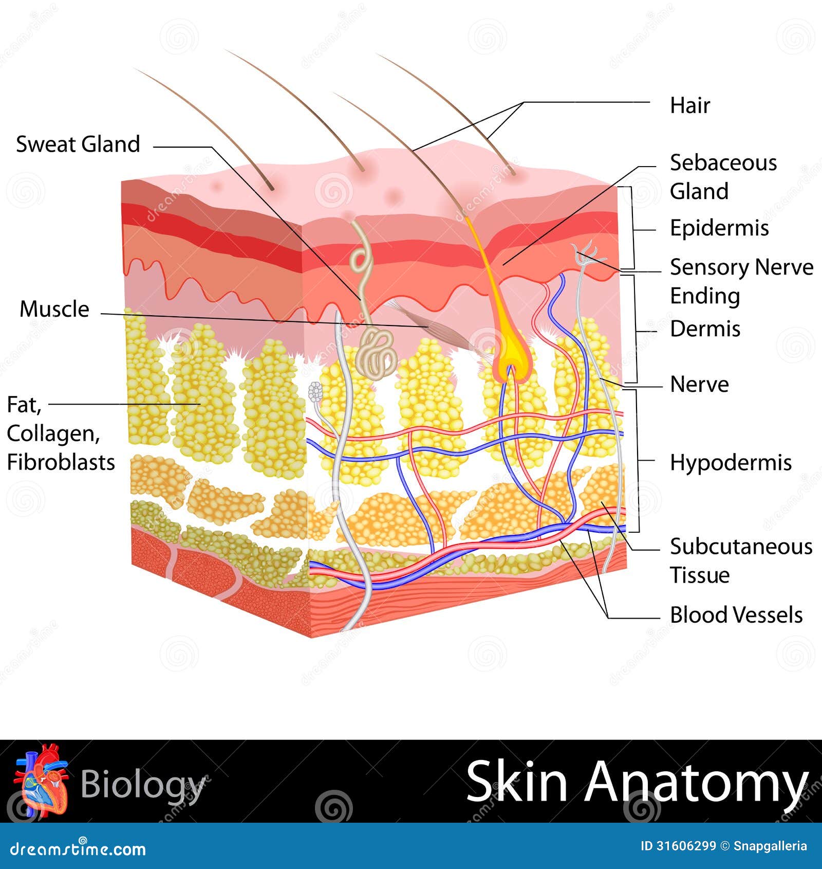
Skin is a waterproof, flexible, but tough protective covering for your body. Normally the surface is smooth, punctuated only with hair and pores for sweat. A cross-section of skin shows the major parts. It is divided into three layers. The outer layer is the epidermis. The dermis is in the middle and fat forms the innermost layer.
Integumentary System definition. The integumentary system is a system comprised of organs that are the outermost protective covering of the animal body, the skin, and its various derivatives. The integumentary system protects against many threats such as infection, desiccation, abrasion, chemical assault, and radiation damage.
Skin is the largest organ in the body and covers the body's entire external surface. It is made up of three layers, the epidermis, dermis, and the hypodermis, all three of which vary significantly in their anatomy and function. The skin's structure is made up of an intricate network which serves as the body's initial barrier against pathogens, UV light, and chemicals, and mechanical injury.
Quiz: Label The Parts Of The Eye. People say that the eyes are the windows to a person's soul. In the class today, we covered parts of the eye, and what changes in them should be alarming to a patient. How much did you get to understand about the human eye?
The skin has three basic layers — the epidermis, the dermis, and the hypodermis. Epidermis. The epidermis is the outermost layer. It is a waterproof barrier that gives skin its tone.
A Skin Diagram Coloring and Labeling Worksheet is a great way to help you keep track of your supplies, materials, and supplies in the industry. These worksheets can be easy to make and they're also easy to copy so you can print them at home. This worksheet can easily be used by the complete team.
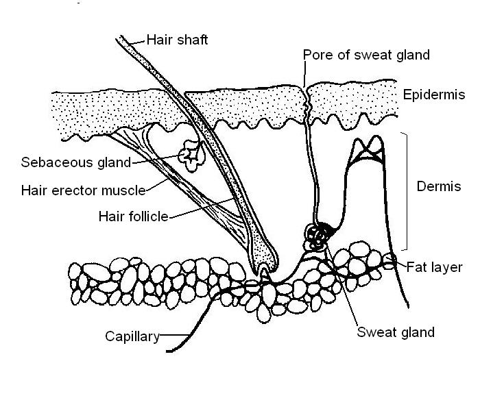
Hair follicles are tiny holes or pores in your skin. Their main function is to grow hair. The scalp of your head too has hair follicles. In biological terms, hair follicle looks like a tunnel-shaped structure situated in the epidermis (outer layer of the skin) . Hair growth starts at the bottom of the hair follicle.
Feb 1, 2011 — Making lava fudge is a great holiday activity (and could make great gifts) – use it to help kids learn about the different proportions of ...
Question. Draw a labelled diagram to show the metaphase stage of mitosis in an animal cell having '6' chromosomes. Ans. The diagram below represents a stage during cell division. Study the same and then answer the questions that follow: Question. Name the parts labelled 1, 2 and 3. Ans. 1= Centromere 2= Spindle fibres 3= Chromatid. Question.
The integumentary system is made up of several organs and structures including the skin, hair, nails, glands, and nerves. The primary function of the integumentary system is to protect the inside of the body from elements in the environment—like bacteria, pollution, and UV rays from the sun. The skin and its associated structures also retain ...
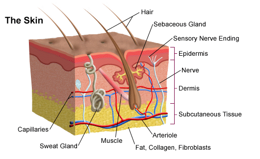
Labeled diagram of the skin. So what's the idea? Spend some time analyzing the skin diagram labeled above. Try to memorize the appearance and location of each structure. Learning the function of each structure will accelerate your ability to memorize, so be sure to check out our detailed article on The Integumentary System parts and functions ...
by S Lawton · 2019 · Cited by 19 — Structure of the skin ... The skin is divided into several layers, as shown in Fig 1. The epidermis is composed mainly of keratinocytes. Beneath ...
Skin label diagram diagram | quizlet
Labeled diagram of the external genital organs that make up the vulva. ... are the two skin folds that lie between the labia majora and the vaginal opening. In contrast to the labia majora, the ...

Cross section of human skin with labels. | canstock
Aug 1, 2021 — WebMD's Skin Anatomy Page provides a detailed image of the skin and its ... Antiviral drugs: Medicines can suppress the activity of the ...
Human skin hair structure anatomical sign stock vector ...
Muscular system structure of muscles. These graphics inspire confidence in any exam room, waiting area, lab area. Superficial back muscles, intermediate back the intrinsic muscles are named as such because their embryological development begins in the back system diagram labeled 209 human muscular system diagram labeled.

30 label the skin structures - label design ideas 2020
Lymphatic system (anterior view) The lymphatic system is a system of specialized vessels and organs whose main function is to return the lymph from the tissues back into the bloodstream.. Lymphatic system is considered as a part of both the circulatory and immune systems, as well as a usually neglected part of students' books. The functions of the lymphatic system complement the bloodstream ...

Associate degree nursing physiology review | skin anatomy ...
Aug 8, 2019 - Label The Skin Anatomy Diagram Tag Human Skin Diagram Label Human Anatomy Diagra.

Structure and function of skin | biology for majors ii
a-c Label-free RCM images of three different types of ex vivo skin tissue areas, including a normal skin, b a melanocytic nevus, and c skin containing BCC, which are used as input of the virtual ...
Skin anatomy stock illustrations – 15,665 skin anatomy stock ...
Start studying Skin Structure labeling. Learn vocabulary, terms, and more with flashcards, games, and other study tools. Rating: 3 · 2 reviews

Quiz 1 - basic skin structure (1 point each)
Onion Cells Under The Microscope Requirements Preparation And. Sketch The Onion Peel Cell As Seen Under The Microscope Label The. Lm Of Onion Skin Stock Image C012 1141 Science Photo Library. Solved 1 Hold A Piece Of Onion And Break In Such A Way T. Onion Cells Under Microscope Stock Footage Video 100 Royalty.
Skin: labeling diagram,review / research worksheets| digital ...
Gland Diagram In this activity, students are going to create a poster that compares the two types of glands found in the skin that are described in the lesson, sweat glands and sebaceous glands.

Skin structure labeling diagram | quizlet
Read the definitions, then label the skin anatomy diagram below. blood vessels - Tubes that carry blood as it circulates. Arteries bring oxygenated blood from ...
Label skin diagram printout - enchantedlearning.com
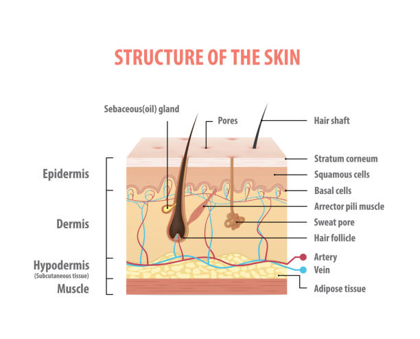
Figure 1. The skin is composed of two main layers: the epidermis, made of closely packed epithelial cells, and the dermis, made of dense, irregular connective ...

Human skin - wikipedia
Figure: Labeled diagram of plant cell, created with biorender.com. The typical characteristics that define the plant cell include cellulose, hemicellulose and pectin, plastids which play a major role in photosynthesis and storage of starch, large vacuoles responsible for regulating the cell turgor pressure.
Solved: label the diagram of human skin below. | chegg.com
The non-keratinized stratified squamous epithelium is a type of squamous epithelium in which the cells are moistened by mucus from the salivary or mucus glands rather than keratin deposits. The nuclei and most metabolic functions of the flattened cells of the surface layer are preserved in this circumstance.
Anatomy of the skin
The 3 meningeal layers are labeled with the stars. The outermost layer of the meninges is the dura mater, which is located beneath the skull. Below the dura mater is the arachnoid, which is the middle meningeal layer. There is a space below the arachnoid called the subarachnoid space, and this is where the CSF is located.

Skin anatomy cross section with labels on white stock photo ...
Body tube/Head. It is the structure that connects the eyepiece to the lenses. Image 2: The body tube part of a microscope is where the ray of light is bent to allow the object being viewed to enlarge by the scope. Picture Source: slideplayer.com. 3. Turret/Nose piece. It is the revolving part of the microscope.
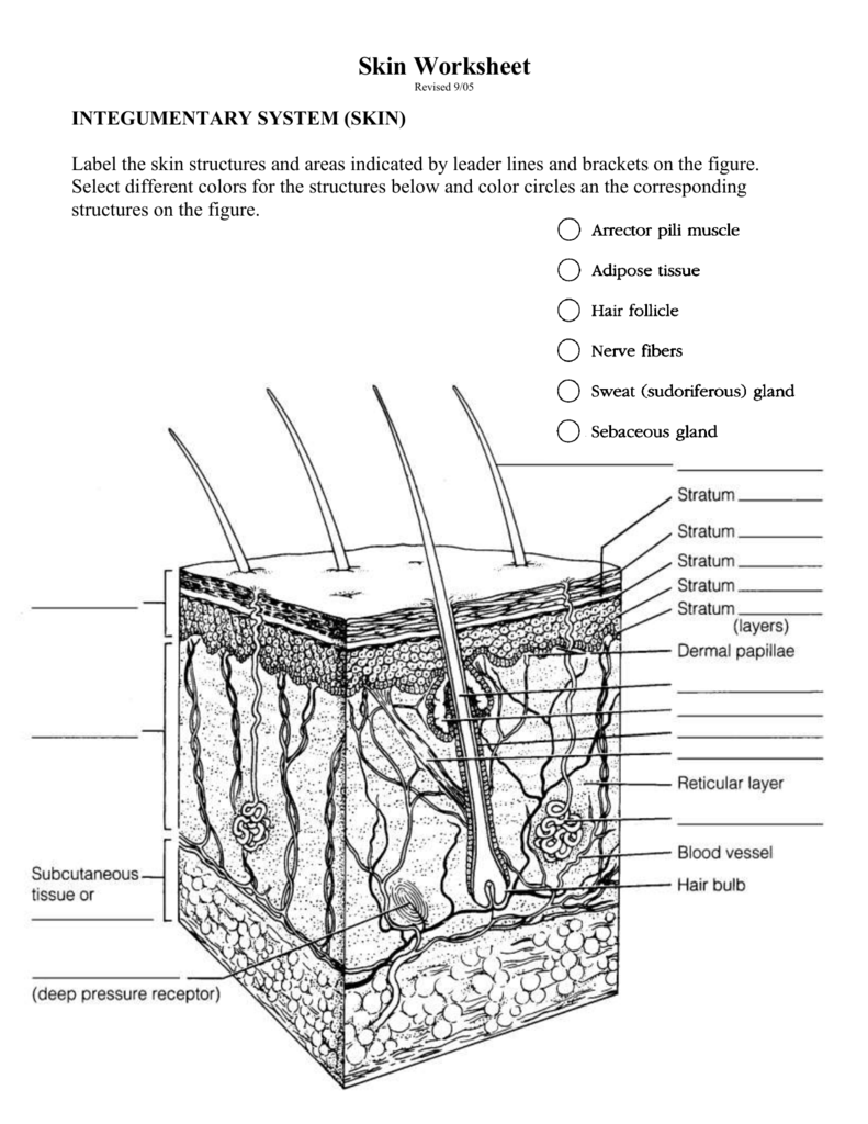
Skin worksheet
Labeled cross-sectional anatomy of the mouse on micro-CT. These images of a normal female Swiss mouse have been acquired with a laboratory-based microCT system (nanoScan PET/CT Mediso (Budapest, Hungary) with an operation voltage of 50kVp and a 0,14mm pitch, with an intravenous injection of 2ml of Visipaque (320mg d'I/ml), at CERIMED (Centre Européen de Recherche en Imagerie Médicale ...

A diagrammatic representation of the structure of human skin ...
7,584 skin diagram stock photos, vectors, and illustrations are available royalty-free. See skin diagram stock video clips. of 76. skin, structure skin aging stages wrinkle skin structure of the skin the skin anatomy needle skin structure skin collagen infographic skin wrinkles skin glands. Try these curated collections. Search for "skin ...
Skin diagram to label - labelled diagram
Skin is part of the integumentary system and considered to be the largest organ of the human body. There are three main layers of skin: the epidermis, the dermis, and the hypodermis (subcutaneous fat). The focus of this topic is on the epidermal and dermal layers of skin. Skin appendages such as sweat glands, hair follicles, and sebaceous glands are reviewed in-depth elsewhere.[1]
Untitled
Draw a neat diagram of the stomatal apparatus found in the epidermis of leaves and label the Stoma, Guard cells, Chloroplast, Epidermal Cells, cell wall and Nucleus. asked Jan 2, 2019 in Class X Science by navnit40 Expert ( 40.5k points)

Skin labeling quiz
19,037 skin anatomy stock photos, pictures & royalty-free ...

Solved exercise 1: skin anatomy label the blank lines in ...

The typical structure of mammalian skin. | download ...
31 label the skin structures and areas indicated in the ...

Skin appendages hair nails glands september 23 24
The anatomy and physiology of animals/skin worksheet ...

Integumentary system labeling diagram | quizlet

33 label the structure of the skin - label design ideas 2020

Skin diagram to label - google search | skin structure, skin ...
Integumentary system: definition, diagram and function | kenhub
A human body skin-structure quiz! - proprofs quiz

Screen shot 2020-08-26 at 12.21.00 am.png - 4 label the skin ...
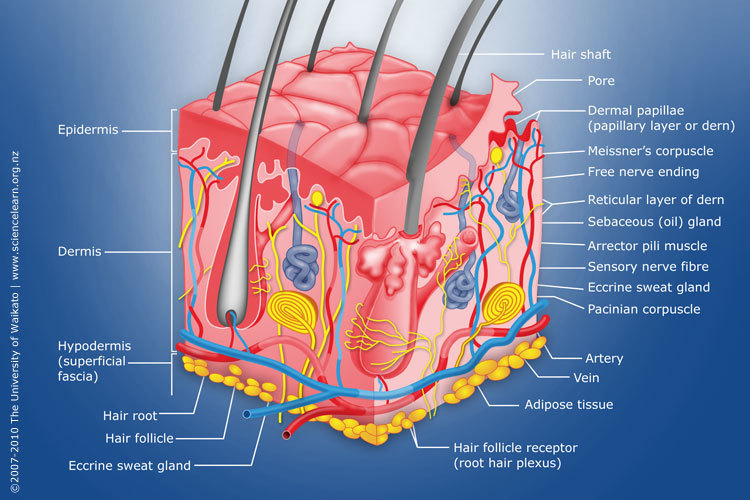
Diagram of human skin structure — science learning hub
0 Response to "34 skin structure diagram to label"
Post a Comment