40 gram negative bacteria diagram
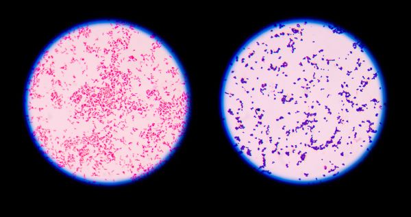
A Gram Stain of a Mixture of Gram-Positive and Gram-Negative Bacteria. Note Gram-negative (pink) bacilli and Gram-positive (purple) cocci. acid-fast Bacteria : These resist decolorization with an acid-alcohol mixture during the acid-fast stain procedure, retain the initial dye carbolfuchsin and appear red when observed through the microscope. Compare and contrast the cell walls of typical Gram-positive and Gram-negative bacteria. 3. Relate bacterial cell wall structure to the Gram-staining reaction. 37 . 38 Bacterial Cell Wall • Peptidoglycan (murein) -rigid structure that lies just outside the cell plasma membrane
Christian Gram, a Danish Physician in 1884 developed a staining technique to distinguish two types of bacteria. The two categories of bacteria based on gram staining are Gram positive bacteria and Gram negative bacteria. Bacteria are first stained with crystal violet or gentian violet.

Gram negative bacteria diagram
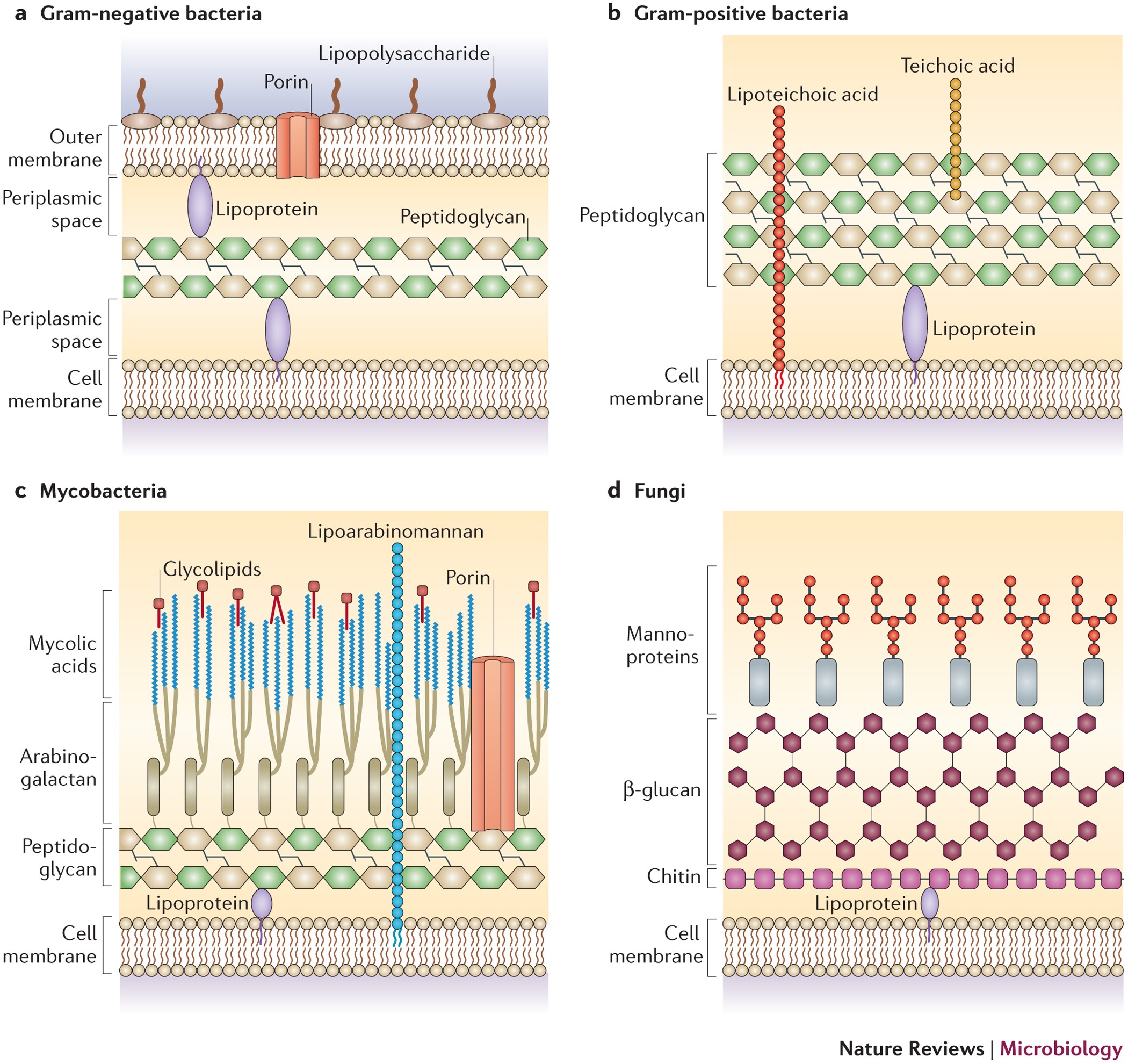
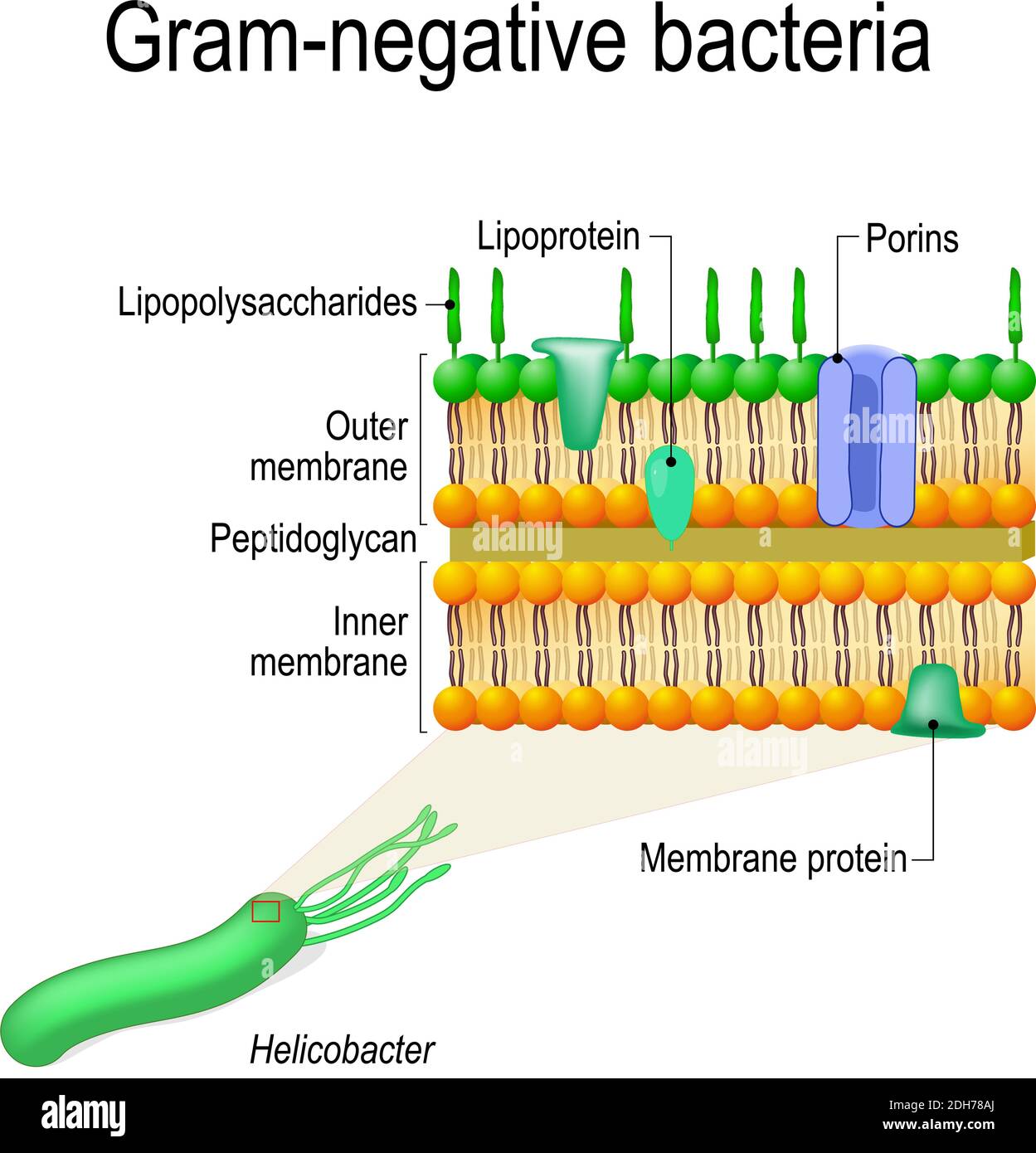
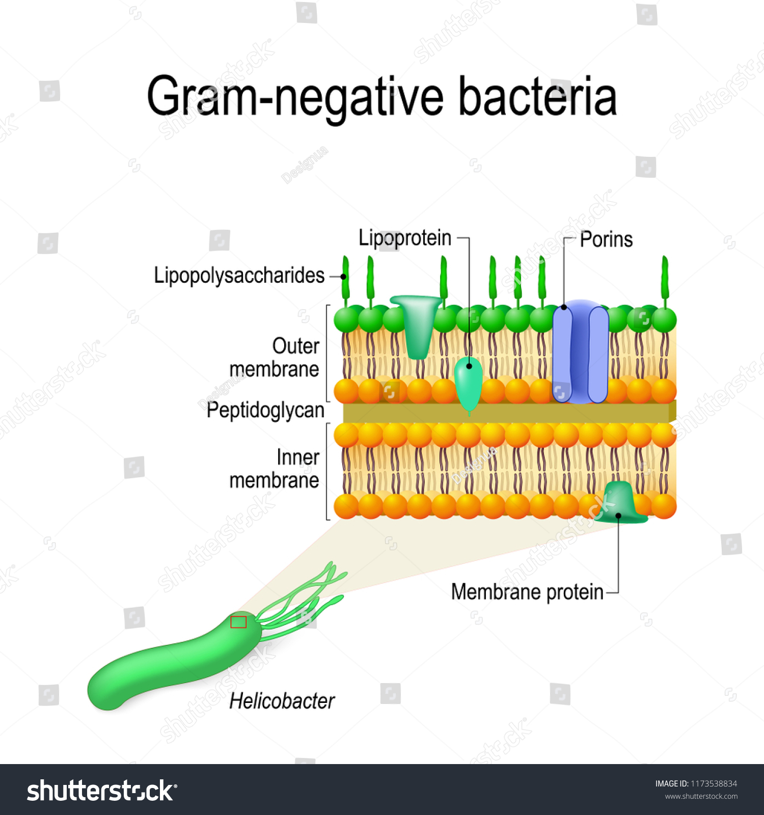
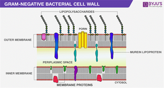
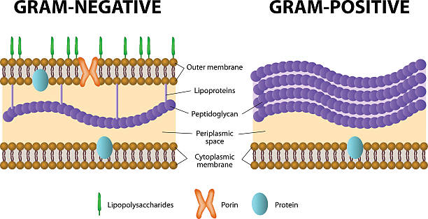
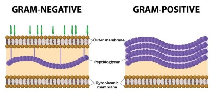
Gram-negative bacteria cell wall. The Cell wall of the Gram-Negative Bacteria is very complex as compared to that of Gram-Positive Bacteria. Combined with the major role of the outer membrane of the cell, with a layer of peptidoglycan, its functional properties are complex, and here is a description of the cell wall and its functional parts. Gram-negative bacteria display these characteristics: . An inner cell membrane is present (cytoplasmic); A thin peptidoglycan layer is present (this is much thicker in gram-positive bacteria); Has outer membrane containing lipopolysaccharides (LPS, which consists of lipid A, core polysaccharide, and O antigen) in its outer leaflet and phospholipids in the inner leaflet 1. cover different classification schemes for grouping bacteria, especially the use of the Gram stain 2. describe the different types of bacteria 3. discuss bacterial structure and the function of the different bacterial components 4. discuss the distinguishing characteristics of Gram positive and Gram negative bacteria.
Gram negative bacteria diagram. The gram-positive bacteria retain the crystal violet colour and stains purple whereas the gram-negative bacteria lose crystal violet and stain red. Thus, the two types of bacteria are distinguished by gram staining. Gram-negative bacteria are more resistant against antibodies because their cell wall is impenetrable. Gram Negative Flow Chart. 13a Gram Negative Bacteria Flashcards Quizlet. Flowchart For Identification Of Anaerobic Gram Positive Bacilli 1 Scientific Diagram. Gram Negative Bacteria Positive Microbiology Flowchart Png 1000x702px Gramnegative Anaerobic Anism Area Bacillus. Solved When Scientific Try To Identify And Unknown Bacter Chegg. Gram Negative Bacteria. Like Gram positive bacteria, Gram negative bacteria are also well distributed in different environments across the globe. Whereas E. coli among other Gram negative bacteria can be found in the gastrointestinal tract, a number of species can be found in marine environments. Start studying Microbiology: Compare and contrast gram-positive and gram-negative bacteria. Learn vocabulary, terms, and more with flashcards, games, and other study tools.
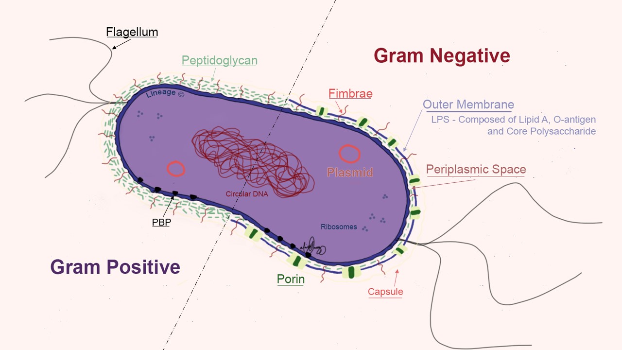
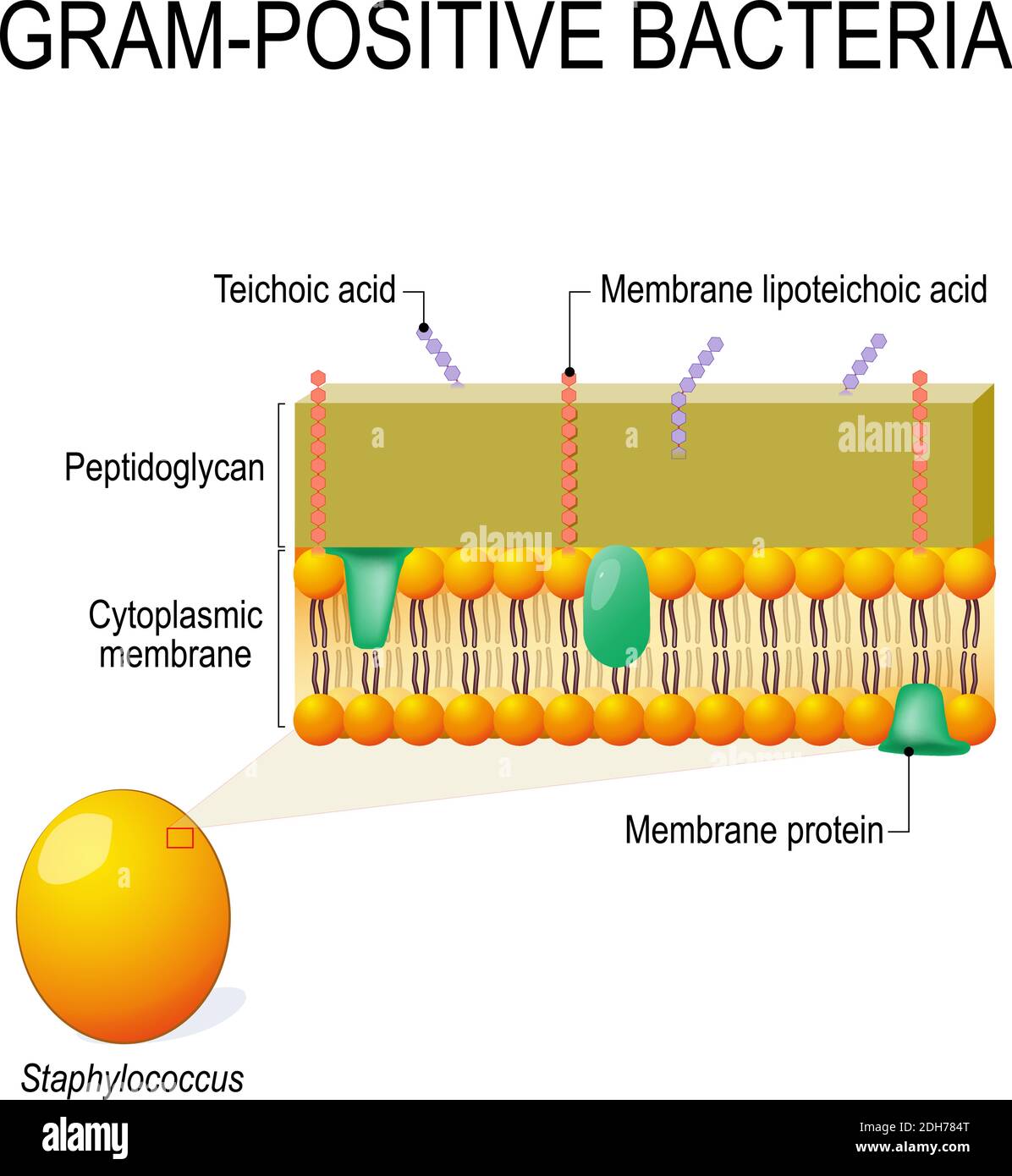
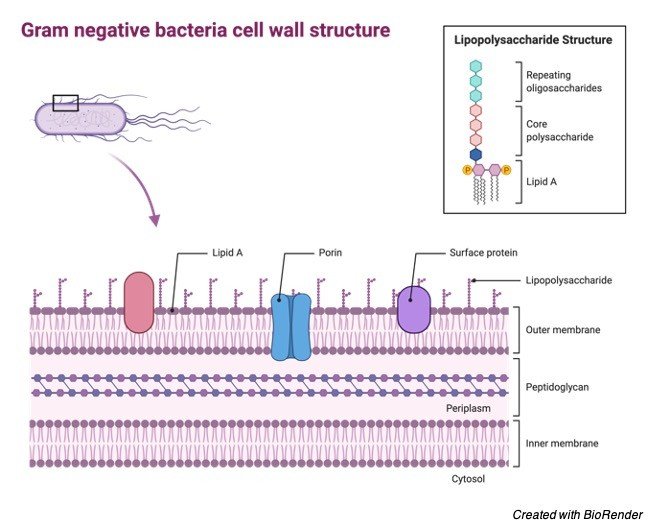
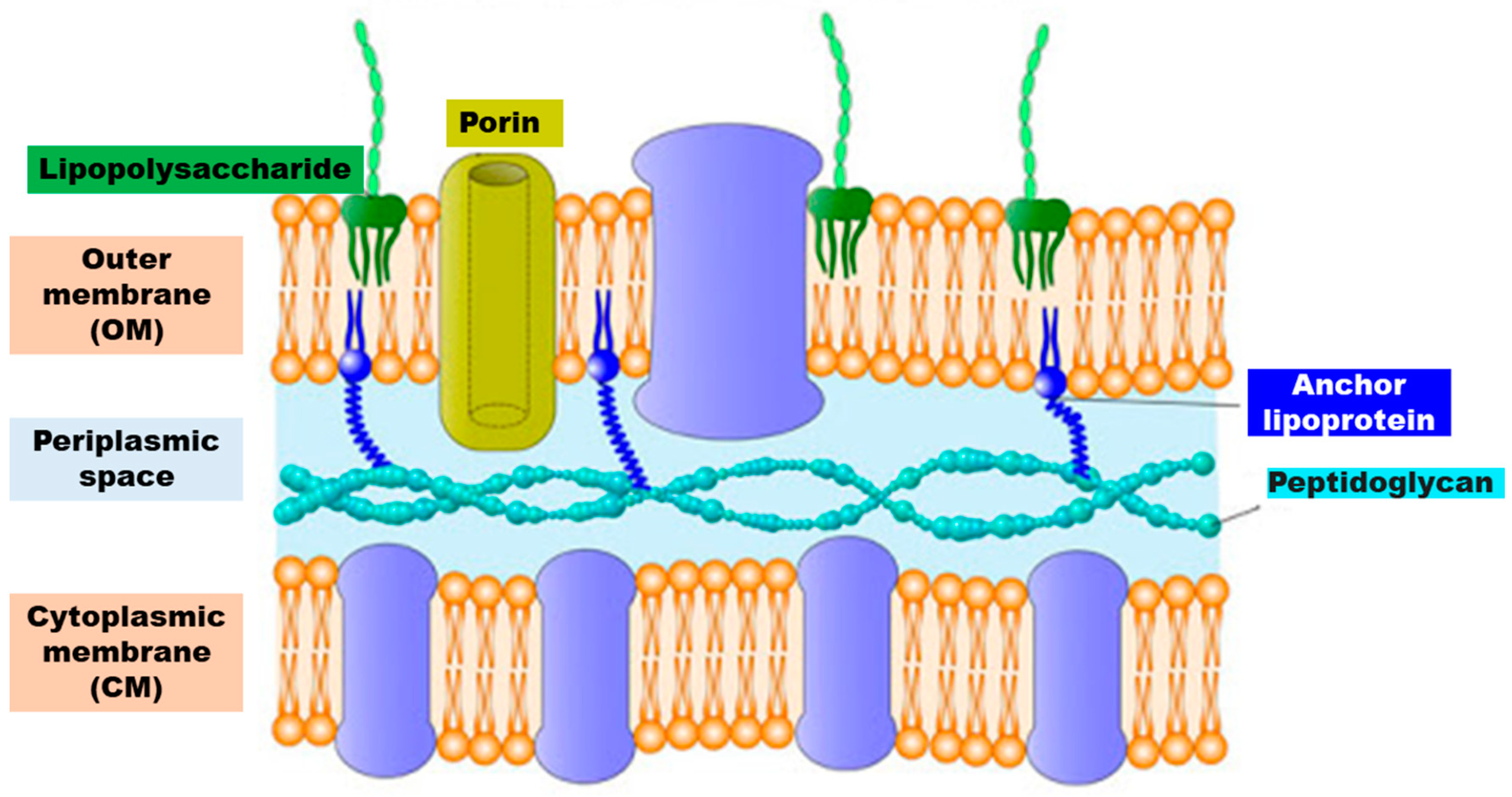
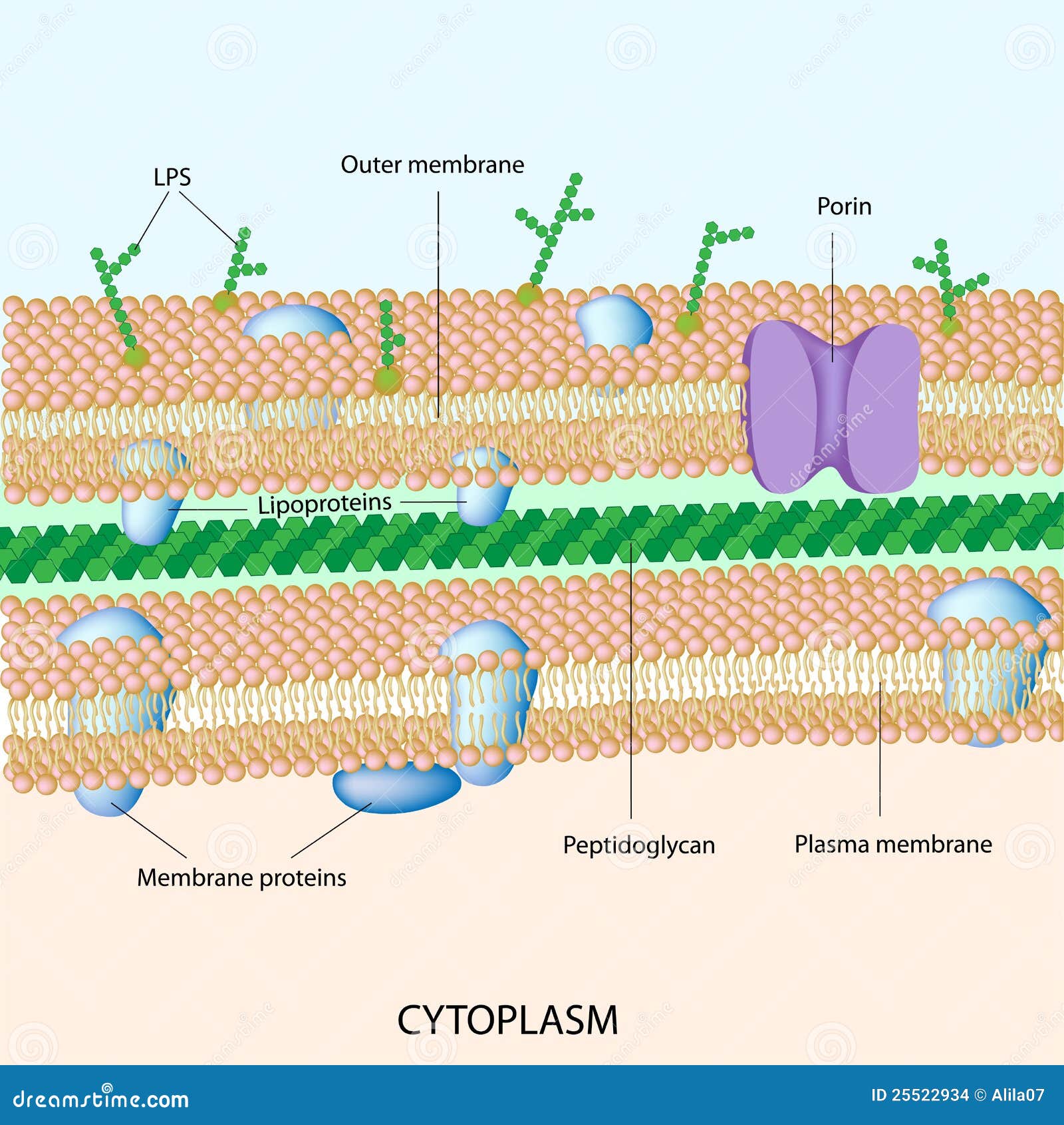
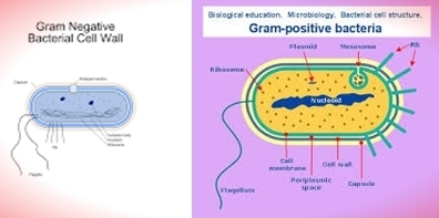
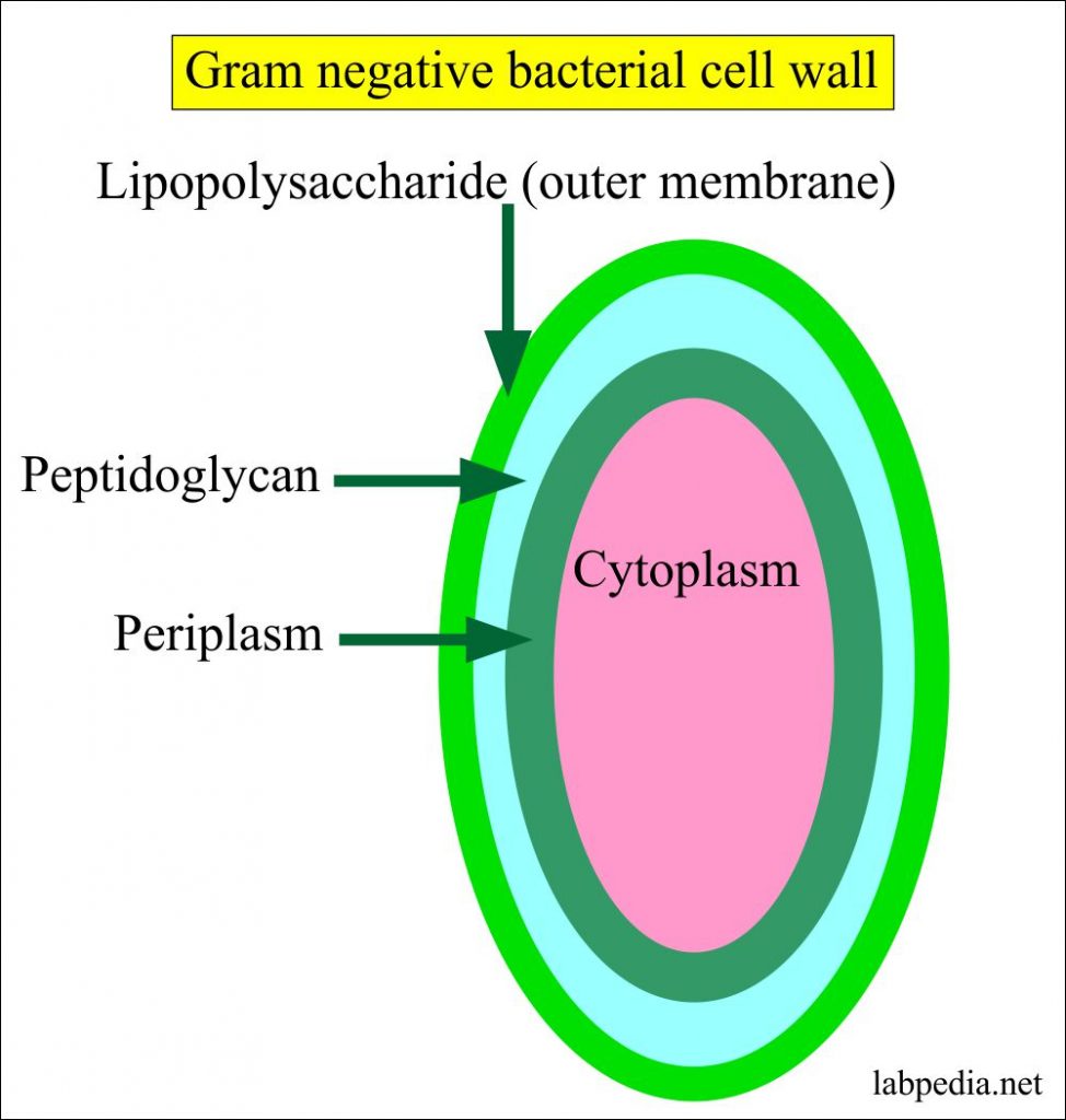
The cell wall structure of Gram negative bacteria is more complex than that of Gram positive bacteria. Located between the plasma membrane and the thin peptidoglycan layer is a gel-like matrix called periplasmic space. Unlike in Gram positive bacteria, Gram negative bacteria have an outer membrane layer that is external to the peptidoglycan ... Mycoplasma are bacteria that have no cell wall and therefore have no definite shape. Outer Membrane: This lipid bilayer is found in Gram negative bacteria and is the location of lipopolysaccharide (LPS) in these bacteria. Gram positive bacteria lack this layer. LPS can be toxic to a host and can stimulate the host's immune system. Laboratory. Flowchart for identification of anaerobic gram positive bacilli 1 scientific diagram diffe size shape and arrangement of bacterial cells solved need help making a flow chart for my gram negative chegg evaluation of two rapid molecular test systems to elish an algorithm for fast identification bacterial pathogens from positive blood ... The diagram below illustrates the differences in the structure of Gram positive and Gram negative bacteria. The two key features that lead to the differing visualization properties of Gram positive and Gram negative species are the thickness of the peptidoglycan layer and presence or absence of the outer lipid membrane.
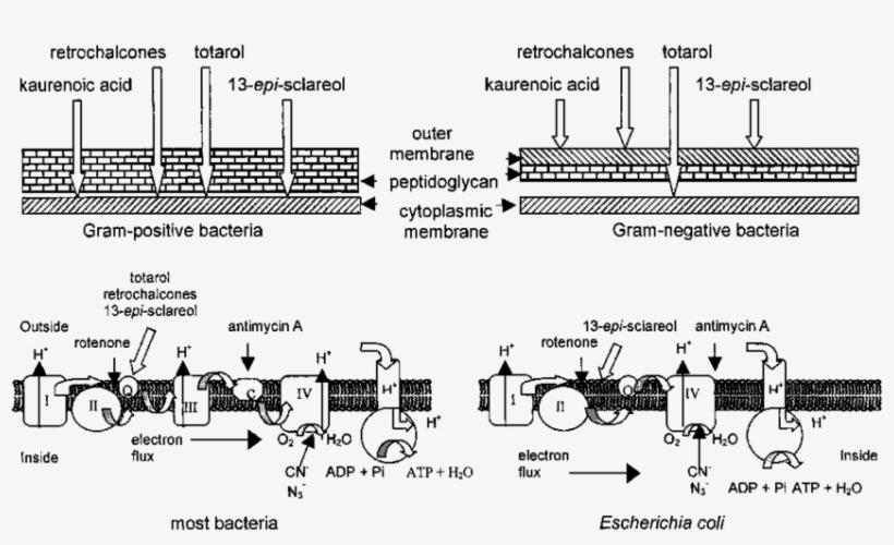
Gram-negative bacteria have a smaller amount of this rigid structure than do gram-positive bacteria. Periplasmic space. an open area between the cell wall and cell membrane in the cell envelopes of bacteria. gram negative bacteria have a more extensive space than do a gram positive bacteria. Cell membrane. Gram-negative cell walls are strong enough to withstand ∼3 atm of turgor pressure (), tough enough to endure extreme temperatures and pHs (e.g., Thiobacillus ferrooxidans grows at a pH of ≈1.5) and elastic enough to be capable of expanding several times their normal surface area ().Strong, tough, and elastic … the gram-negative cell wall is a remarkable structure which protects the ... Experts are tested by Chegg as specialists in their subject area. We review their content and use your feedback to keep the quality high. 100% (4 ratings) Transcribed image text: Identify the Gram-negative and Gram-positive bacteria and their flagellum components on the diagram below. Gram-negative Gram-positive Hook Filament Basal body. Electron Transport Chain of Bacteria (With Diagram) The electron transport chains of bacteria (prokaryotes) operate in plasma membrane (mitochondria are absent in prokaryotes). Some bacterial electron transport chains resemble the mitochondrial electron transport chain. Paracoccus denitrificans is a gram-negative, facultative anaerobic soil ...

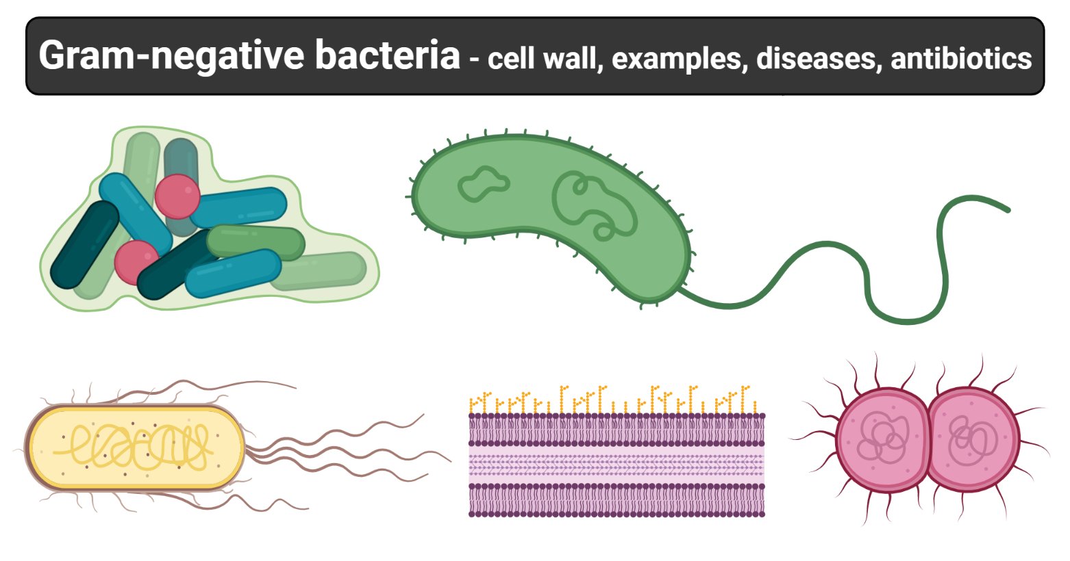
Klebsiella, Enterobacter, and Serratia are closely related gram-negative bacteria Overview of Gram-Negative Bacteria Gram-negative bacteria are classified by the color they turn after a chemical process called Gram staining is used on them. Gram-negative bacteria stain red when this process is used.

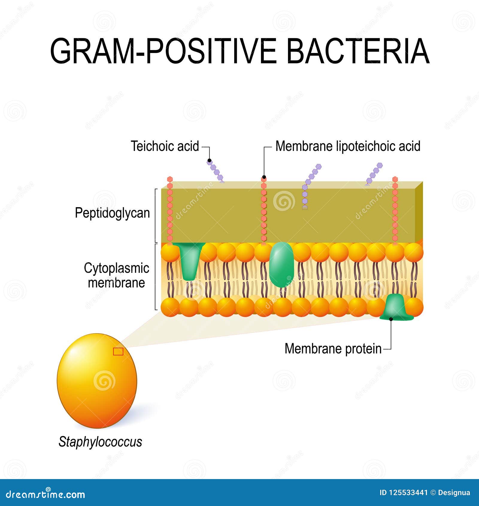
Gram-negative Bacteria. Go to PAGE 2 > Gram-positive Cells. In Gram-positive bacteria, peptidoglycan makes up as much as 90% of the thick cell wall enclosing the plasma membrane. See Page 2 for a diagram of the Gram-negative cell wall and a video on ...

The gram stain procedure distinguishes between gram positive and gram negative groups by coloring these cells red or violet. The diagram below illustrates the differences in the structure of gram positive and gram negative bacteria. Most of the gram positive cell wall contain considerable amount of teichoic acid and teichuronic acid.
Gram negative bacteria can be either cocci or bacilli. Gram negative pathogenic bacteria commonly encountered are E.coli, Klebsiella, Salmonella spp, shigella, etc 2. Albert staining: is performed in case if one suspects a Corynebacterium spp. 3. Acid fast staining: is performed in cases suspected of Mycobacterial
In segment 13 i.e. Toxin production it is more accurate to write under gram negative bacteria exotoxins and/or endotoxins rather than exotoxins or endotoxins because endotoxins are produced by all gram negative bacteria as it ( the LPS) is an integral part of the outer membrane, so any species produce exotoxins will already produce both.

We will now look at the Gram-negative bacterial cell wall. As mentioned in the previous section on peptidoglycan, Gram-negative bacteria are those that decolorize during the Gram stain procedure, pick up the counterstain safranin, and appear pink (Figure 2.3 B. 2 B.1). Figure 2.3 B. 2 B.1: Gram Stain of Escherichia coli.
The cell wall of Gram negative bacteria is thin and is composed of peptidoglycan.; The cell envelope has 3 layers including, a unique outer membrane, a thin peptidoglycan layer, and the cytoplasmic membrane. An outer membrane of the cell wall is a bilayer structure consisting of phospholipids molecules, lipopolysaccharides (LPS), lipoproteins and surface proteins.
Flagella occur on both Gram-positive and Gram-negative bacteria, and their presence can be useful in identification. For example, they are found on many species of bacilli but rarely on cocci. In contrast, pili occur almost exclusively on Gram-negative bacteria and are found on only a few Gram-positive organisms (e.g., Corynebacterium renale).
Medical Relevance of Gram Negative Cell Wall: The cell wall of Gram-negative bacteria is often a virulence factor that enables pathogenic bacteria to cause disease. The virulence of Gram-negative bacteria is often associated with certain components of the cell wall, in particular, the lipopolysaccharide ( otherwise known as LPS or endotoxin).

Identification Flow Chart For Isolate D9 Supporting Figures On Gram Scientific Diagram. Solved What Two Broups Of Gram Negative Bacteria Can Be Diffeiated With The Oxidase Test Results Whyd 2 Points You Are Trying To Identify A Known Positive Bacterial Species Mycobacterium Tis Withthe Help.
Gram stain and bacterial morphology: Of all the different classification systems, the Gram stain has withstood the test of time. Discovered by H.C. Gram in 1884 it remains an important and useful technique to this day. It allows a large proportion of clinically important bacteria to be classified as either Gram positive or negative based on their

1. cover different classification schemes for grouping bacteria, especially the use of the Gram stain 2. describe the different types of bacteria 3. discuss bacterial structure and the function of the different bacterial components 4. discuss the distinguishing characteristics of Gram positive and Gram negative bacteria.
Gram-negative bacteria display these characteristics: . An inner cell membrane is present (cytoplasmic); A thin peptidoglycan layer is present (this is much thicker in gram-positive bacteria); Has outer membrane containing lipopolysaccharides (LPS, which consists of lipid A, core polysaccharide, and O antigen) in its outer leaflet and phospholipids in the inner leaflet
Gram-negative bacteria cell wall. The Cell wall of the Gram-Negative Bacteria is very complex as compared to that of Gram-Positive Bacteria. Combined with the major role of the outer membrane of the cell, with a layer of peptidoglycan, its functional properties are complex, and here is a description of the cell wall and its functional parts.


0 Response to "40 gram negative bacteria diagram"
Post a Comment