38 pulmonary embolism pathophysiology diagram
by DC Sabiston Jr · 1965 · Cited by 63 — Diagrammatic illustration of thrombosis in situ which occurs both proximal and distal to a pulmonary embolus. The proximal thrombosis extends to the first major ... How severe is Pulmonary Embolism? Pulmonary embolism can be grouped based on the location of clot or how sick a person is. Based on location of the clot into pulmonary artery following terms are used A) saddle PE (large clot into main pulmonary artery), B) lobar PE (into big branch of pulmonary artery), or C) distal PE (into small branches of ...
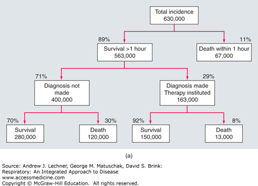
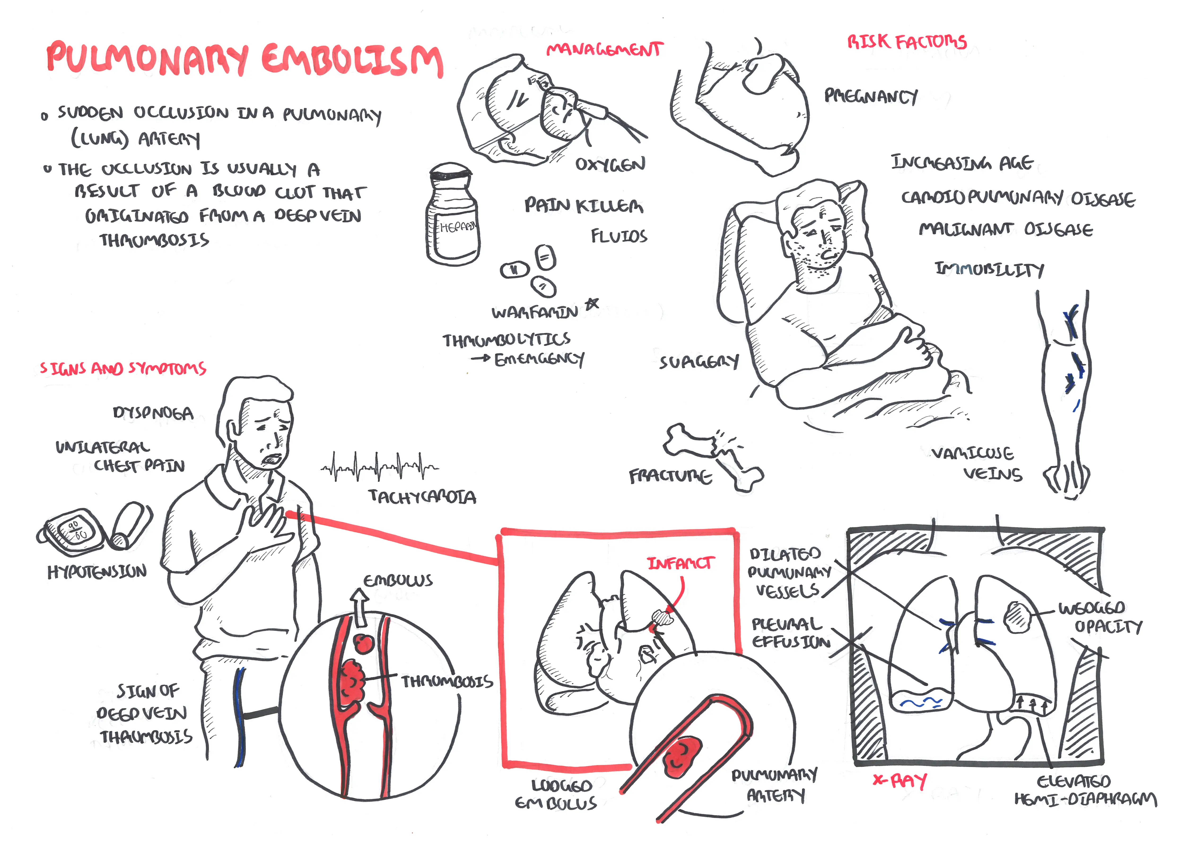
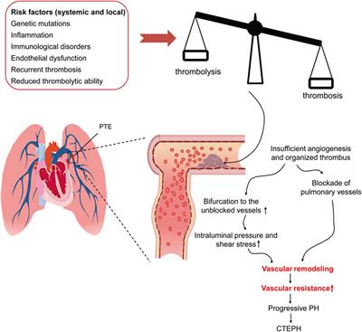
Pulmonary embolism (PE) occurs when there is an acute obstruction of the pulmonary artery or one of its branches. It is commonly caused by a venous thrombus that has dislodged from its site of formation and embolized to the arterial blood supply of one of the lungs. The process of clot formation and embolization is termed thromboembolism.
Pulmonary embolism pathophysiology diagram
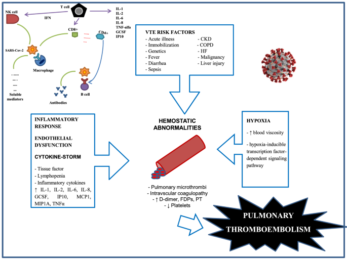
Pulmonary embolus is predominantly due to thrombus breaking off from deep veins or from within the right heart, lodging within large or small vessels within the pulmonary vasculature, causing a variable degree of clinical features ranging from asymptomatic through to shock and cardiac arrest. Non-thrombotic causes include air or fat embolism. Outcome is predicated by the degree of right ... Greenspan RH. Pulmonary angiography and the diagnosis of pulmonary embolism. Prog Cardiovasc Dis. 1994 Sep-Oct; 37 (2):93–105. [Google Scholar] Hull RD, Raskob GE, Ginsberg JS, Panju AA, Brill-Edwards P, Coates G, Pineo GF. A noninvasive strategy for the treatment of patients with suspected pulmonary embolism. Thrombotic pulmonary embolism is not an isolated disease of the chest but a complication of venous thrombosis. Deep venous thrombosis (DVT) and pulmonary embolism are therefore parts of the same process, venous thromboembolism. Evidence of leg DVT is found in about 70% of patients who have sustained a pulmonary embolism; in most of the remainder, it is assumed that the whole thrombus has ...
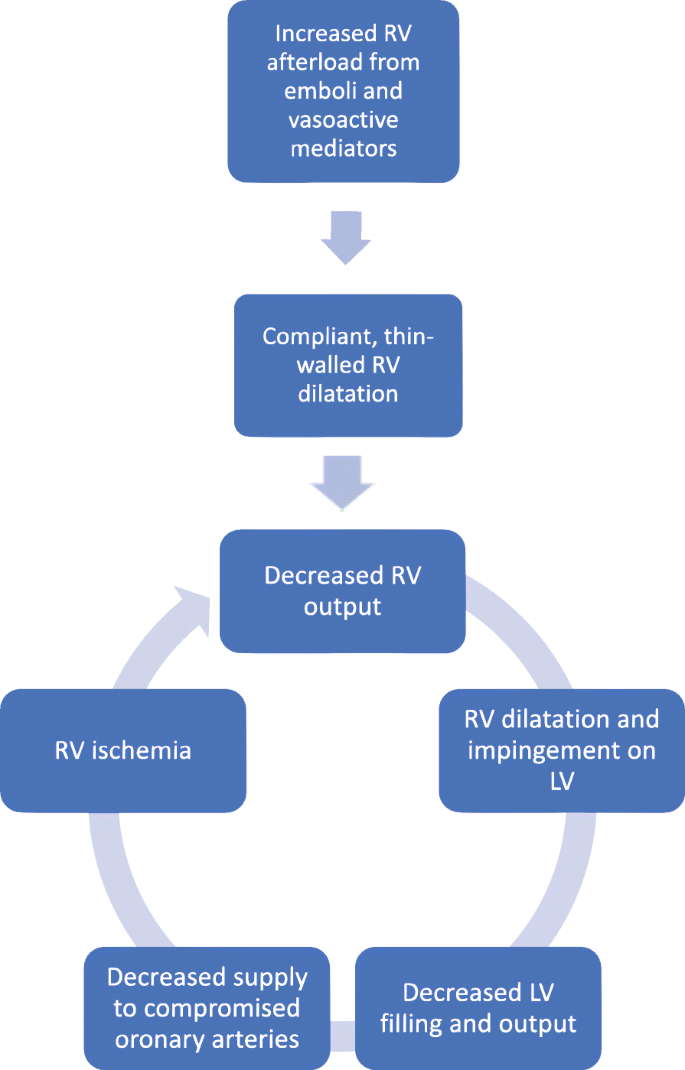
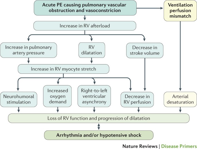
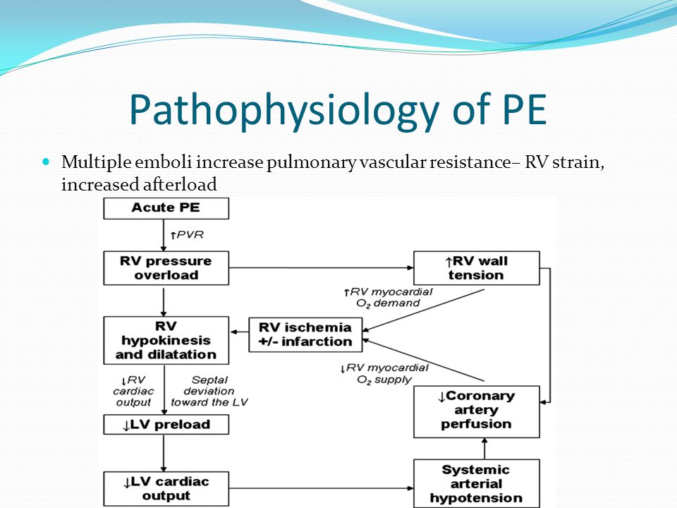
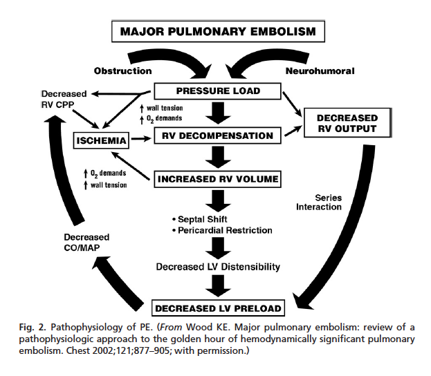
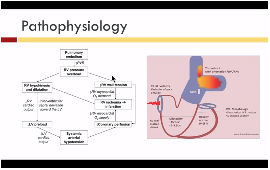
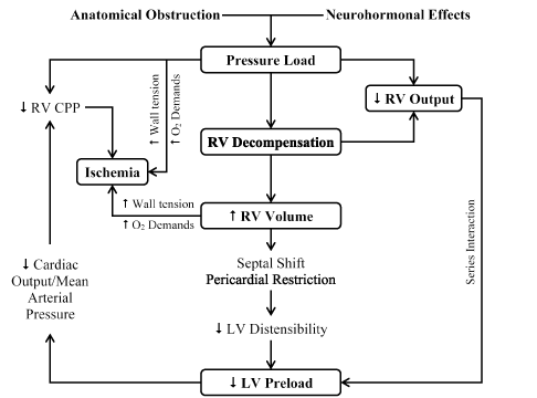
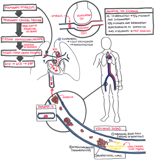
Pulmonary embolism pathophysiology diagram. pulmonary embolism than a patient who was well until the embolic event occurred. Most emboli are multiple. As both the extent and chronicity of obstruction vary so widely, pulmonary embolism can produce widely diVeringclinicalpictures.Disregardingchronic thromboembolic pulmonary hypertension, it is convenient to classify pulmonary embolism 18 Sept 2020 — The pathophysiology of pulmonary embolism. Although pulmonary embolism can arise from anywhere in the body, most commonly it arises from the ... Pulmonary embolism (PE) is responsible for approximately 100,000 to 200,000 deaths in the United States each year. With a diverse range of clinical presentations from asymptomatic to death, diagnosing PE can be challenging. Various resources are available, ... Result in bronchial pulmonary artery constriction. Pulmonary Embolism. Increased pulmonary vascular resistance. Increase in pulmonary arterial Increased right-sided heart workload pressure to maintain pulmonary blood flow. Right sided heart failure. Decreased cardiac output. Decreased BP. Shock. Risk of hypoxemia.
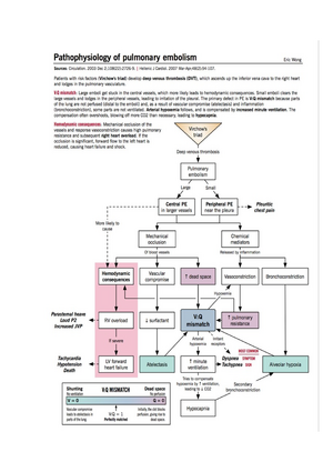
In the first 24 hours, chest x-rays and pulmonary function tests are not definitive for a pulmonary embolism. Oximetry and arterial blood gas typically show hypoxemia. Echocardiography may show right ventricle strain. Serum D-dimer levels will test positive for thrombus degradation by-products; fibrinogen and fibrin. Sep 26, 2012 · Venous thromboembolism (VTE) » Pathophysiology of pulmonary embolism. Venous thromboembolism (VTE) » Pathophysiology of pulmonary embolism. Posted September 26, 2012 by Eric Wong. Pathophysiology of Pulmonary Embolism. Once deep venous thrombosis develops, clots may dislodge and travel through the venous system and the right side of the ... 23 Sept 2019 — Please read and agree to the disclaimer before watching this video.. An embolism is the lodging of an embolus, a blockage-causing piece of ...
by SZ Goldhaber · 2003 · Cited by 474 — This New Frontiers article reviews the epidemiology, pathophysiology, diagnosis, treatment, and prevention of pulmonary embolism (PE) in 2 parts. Thrombotic pulmonary embolism is not an isolated disease of the chest but a complication of venous thrombosis. Deep venous thrombosis (DVT) and pulmonary embolism are therefore parts of the same process, venous thromboembolism. Evidence of leg DVT is found in about 70% of patients who have sustained a pulmonary embolism; in most of the remainder, it is assumed that the whole thrombus has ... Greenspan RH. Pulmonary angiography and the diagnosis of pulmonary embolism. Prog Cardiovasc Dis. 1994 Sep-Oct; 37 (2):93–105. [Google Scholar] Hull RD, Raskob GE, Ginsberg JS, Panju AA, Brill-Edwards P, Coates G, Pineo GF. A noninvasive strategy for the treatment of patients with suspected pulmonary embolism. Pulmonary embolus is predominantly due to thrombus breaking off from deep veins or from within the right heart, lodging within large or small vessels within the pulmonary vasculature, causing a variable degree of clinical features ranging from asymptomatic through to shock and cardiac arrest. Non-thrombotic causes include air or fat embolism. Outcome is predicated by the degree of right ...












0 Response to "38 pulmonary embolism pathophysiology diagram"
Post a Comment