39 g protein coupled receptors diagram
O'Hayre M, Degese MS, Gutkind JS (2014) Novel insights into G protein and G protein-coupled receptor signaling in cancer. Curr. Opin. Cell Biol. 27, 126–35. Pan D (2010) The hippo signaling pathway in development and cancer. Dev. Cell 19(4), 491–505. Sudol M, Harvey KF (2010) Modularity in the Hippo signaling pathway. Trends Biochem. Sci ... G protein-coupled receptors ( GPCRs) are a large family of cell surface receptors that share a common structure and method of signaling. The members of the GPCR family all have seven different protein segments that cross the membrane, and they transmit signals inside the cell through a type of protein called a G protein (more details below).
G-protein coupled receptors are only found in eukaryotes and they comprise of the largest known class of membrane receptors. In fact humans have more than 1,000 known different types of GPCRs, and each one is specific to a particular function. They are a very unique membrane receptor and they are the target of around 30 to 50% of all modern ...

G protein coupled receptors diagram
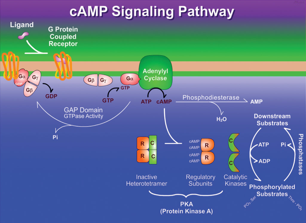
Background The G-protein coupled receptor (GPCR) superfamily consists of structurally similar proteins arranged into families (classes), and is one of the most abundant protein classes in the mammalian genome. 1-5 GPCRs undertake a plethora of essential physiological functions and are targets for numerous novel drugs. 4,5 Their ligands are structurally heterogenous, including natural ... The metabotropic glutamate receptors (mGluRs) are family C G-protein-coupled receptors that participate in the modulation of synaptic transmission and neuronal excitability throughout the central nervous system. The mGluRs bind glutamate within a large ... These receptors are coupled to intracellular GTP-binding proteins (G-proteins). Once activated, G-proteins trigger the production of a variety of second messengers (e.g. cyclic AMP [cAMP], inositol triphosphate [IP3], diacylglycerol [DAG], etc.) helping to regulate a number of body functions ranging from sensation to growth to hormone release.
G protein coupled receptors diagram. G protein coupled receptors and their Signaling Mechanism 1. G Protein Coupled Receptors Faraza Javed Mphil Pharmacology 2. G Protein-Coupled Receptors G protein-coupled receptors (GPCRs), also known as seven-transmembrane domain receptors, 7TM receptors, serpentine receptor, and G protein-linked receptors (GPLR), constitute a large protein family of receptors that sense molecules outside the ... Gustducin is a G protein associated with taste and the gustatory system, found in some taste receptor cells. Research on the discovery and isolation of gustaducin is recent. It is known to play a large role in the transduction of bitter, sweet and umami stimuli. G-Protein Coupled Receptors (GPCRs) Market report 2022 describes detailed study of competitive share analysis, marketing strategies, SWOT analysis, revenue estimates, equipment suppliers, and ... Introduction. G protein-coupled receptors (GPCRs) represent the largest protein family encoded by the human genome. Located on the cell membrane, they transduce extracellular signals into key physiological effects. 1 Their endogenous ligands include odors, hormones, neurotransmitters, chemokines, etc., varying from photons, amines, carbohydrates, lipids, peptides to proteins.
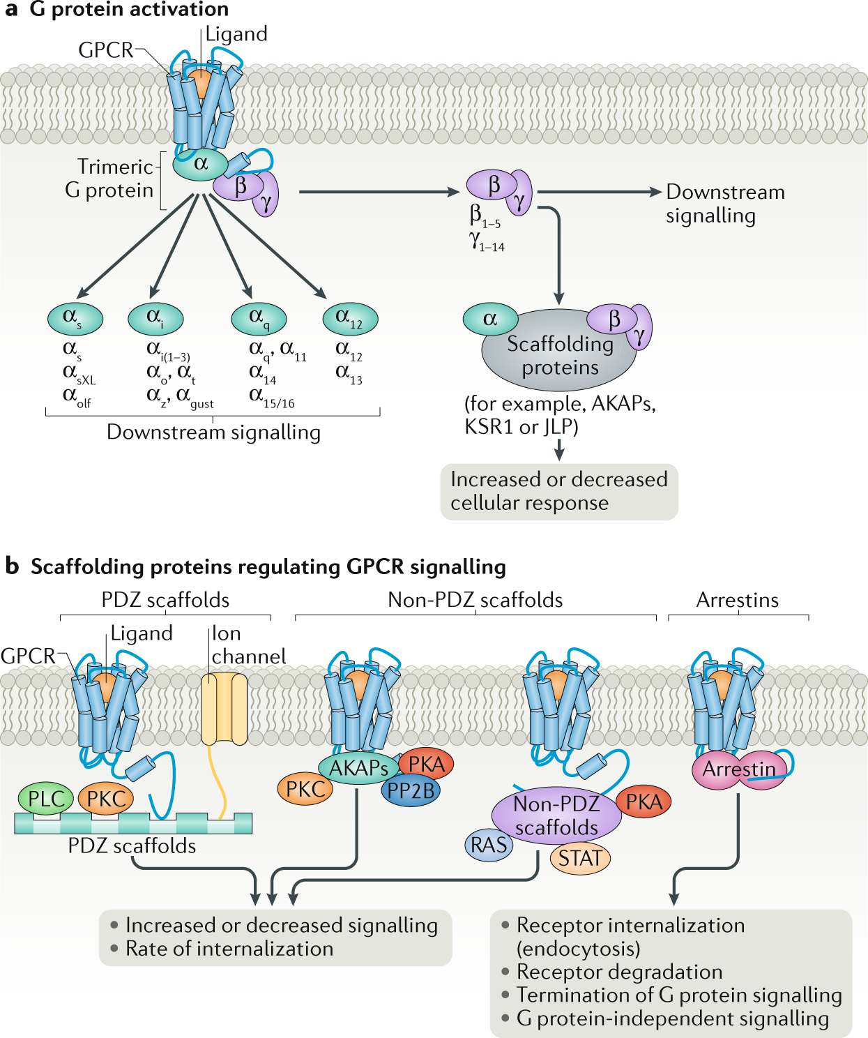
G Protein coupled receptor. the largest family of cell-surface receptors. transmembrane receptors. all very similar in structure. extremely widespread and diverse in their functions. GDP. G Protein Coupled Receptors (GPCRs) regulate a wide variety of normal biological processes and play a role in the pathophysiology of many diseases upon dysregulation of their downstream signaling activities. The intracellular signaling pathways activated by GPCR signaling include cAMP/ PKA pathway, PKC pathway, Ca2+/NFAT pathway, PLC pathway ... Receptor-G protein interactions (chimeras, mutagenesis, peptide blocking etc): 1. efficacy and selectivity of G-protein interactions probably depend on the proper combination of multiple specific cytoplasmic receptor regions 2. residues at cytoplasmic ends of all helices and nearby loops have been implicated for G protein-coupled receptor (GPCR), also called seven-transmembrane receptor or heptahelical receptor, protein located in the cell membrane that binds extracellular substances and transmits signals from these substances to an intracellular molecule called a G protein (guanine nucleotide-binding protein). GPCRs are found in the cell membranes of a wide range of organisms, including mammals ...
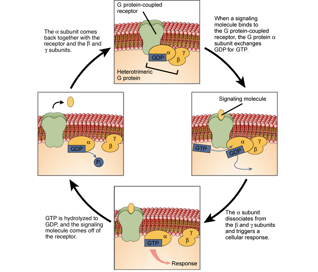
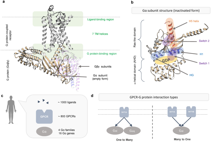
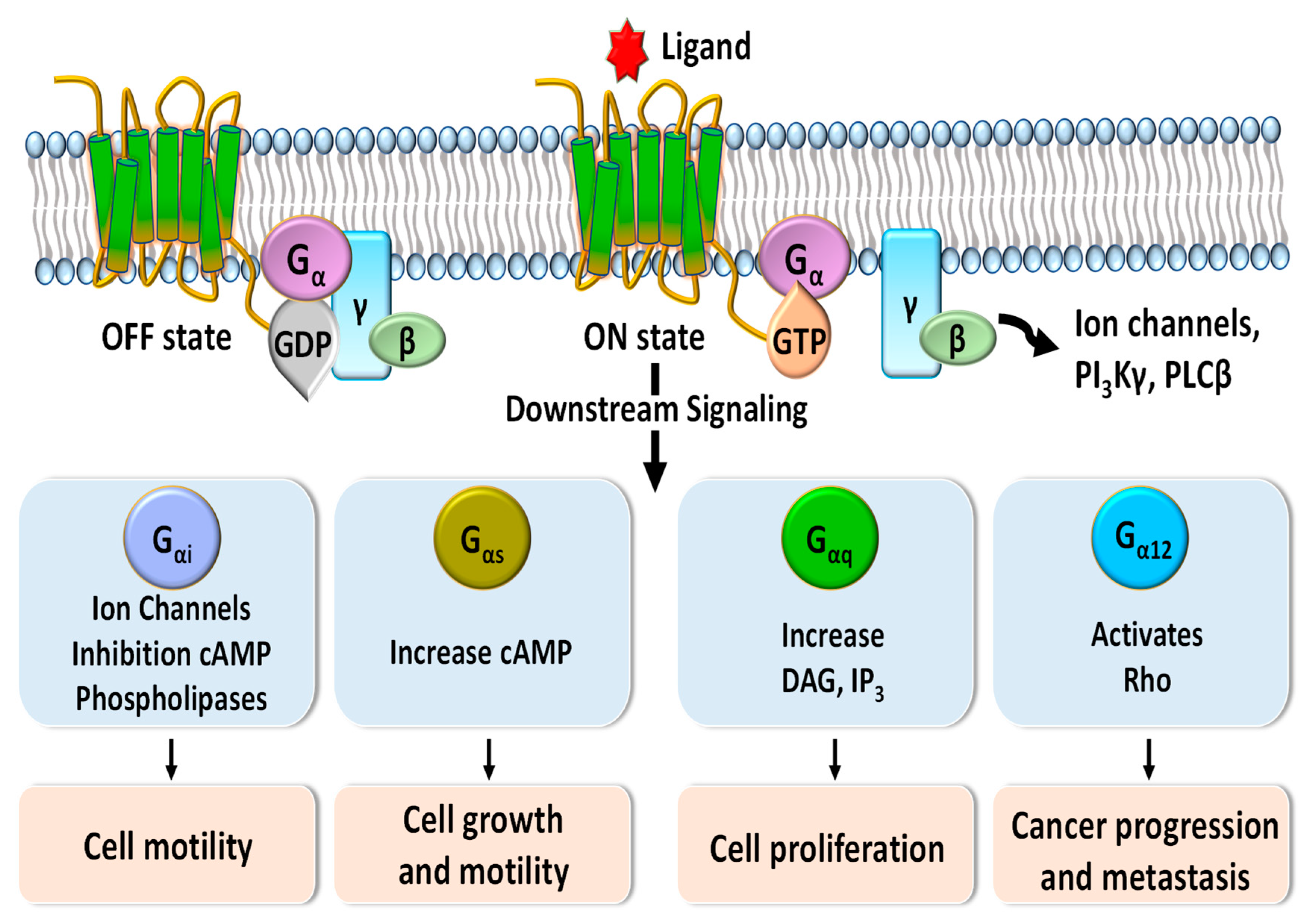
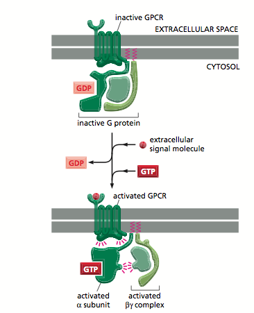
The past two years have seen remarkable advances in the structural biology of G-protein-coupled receptors (GPCRs). Highlights have included solving the first crystal structures of ligand-activated GPCRs—the human β 2 adrenergic receptor (β 2 AR), the avian β 1 AR and the human A 2A adenosine receptor—as well as the structures of opsin and an active form of rhodopsin. Lecture 15 - Functions of G protein coupled receptors in cells-GPCRs fast acting effectors o K+ channels-GPCRs that regulate adenylyl cyclase o cAMP, epithelial cell Cl-channels, PKA, CREB-GPCRs that activate phospholipase C o Ca 2+, IP 3, DAG, PKC, NO, cGMP, PKG-Largest family of receptors: at least 800 G-protein coupled receptors Activation of muscarinic acetylcholine receptor and K ... G protein-coupled receptors (GPCRs) play key roles in intercellular signaling in the brain. Their effects on cellular function have been largely studied in neurons, but their functional consequences on astrocytes are less known. Using both endogenous and chemogenetic approaches with DREADDs, we have … In this diagram of G-protein-coupled receptor activation, the alpha, beta, and gamma subunits are shown with distinct relationships to the plasma membrane. After exchange of GDP with GTP on the...
The G protein-coupled receptor (GPCR) superfamily comprises the largest and most diverse group of proteins in mammals. GPCRs are responsible for every aspect of human biology from vision, taste, sense of smell, sympathetic and parasympathetic nervous functions, metabolism, and immune regulation to reproduction.
The magnocellular neurons in the supraoptic nucleus project to the neural lobe and release vasopressin and oxytocin into the peripheral circulation where they act on the kidney to promote fluid retention or stimulate smooth muscles in the vasculature, ...
28.10.2021 · How two stimuli yield distinct G protein signaling at the same G protein-coupled ... Ribbon diagram of NK1R ... Q. et al. Mini G protein probes for active G protein–coupled receptors (GPCRs ...
What are g-protein coupled receptors? Where are these receptors located in the cell? How do they work? Draw a diagram. 2. What is an antagonist for a receptor? What would an antagonist do? Draw a diagram to explain this. 3. How would caffeine binding to an adenosine receptor affect the activity inside the neuron? Refer to your diagram for ...
23.6.2021 · Various allosteric ligand binding sites, as well as the G-protein (pink) binding site, correspond to essential or hot regions. Complex structure (PDB id: 1pzo) resolved in the presence of two allosteric ligands (spatially neighboring, both shown in magenta sticks) and the orthosteric ligand (yellow sticks).
Antibodies are the secreted form of the B-cell receptor. An antibody is identical to the B-cell receptor of the cell that secretes it except for a small portion of the C-terminus of the heavy-chain constant region. In the case of the B-cell receptor the C-terminus is a hydrophobic membrane-anchoring sequence, and in the case of antibody it is a hydrophilic sequence that allows …
A G protein-coupled receptor (GPCR) ... trigger so many pathways is a key difference between receptor tyrosine kinases and G protein-coupled receptors. 16. Explain what happens in the first step. ! ... 26. This diagram uses testosterone, a hydrophobic hormone, ...
The general function of G q is to activate intracellular signaling pathways in response to activation of cell surface G protein-coupled receptors (GPCRs).GPCRs function as part of a three-component system of receptor-transducer-effector. The transducer in this system is a heterotrimeric G protein, composed of three subunits: a Gα protein such as G αq, and a complex of two tightly linked ...
Binding of histamine to the G-protein coupled histamine H1 receptor plays an important role in the context of allergic reactions; however, no crystal structure of the resulting complex is ...
Adenylyl cyclase (EC 4.6.1.1, also commonly known as adenyl cyclase and adenylate cyclase, abbreviated AC) is an enzyme with key regulatory roles in essentially all cells. It is the most polyphyletic known enzyme: six distinct classes have been described, all catalyzing the same reaction but representing unrelated gene families with no known sequence or structural homology.
GPCR Pathway. G-protein-coupled receptors (GPCRs), also known as seven- (pass)-transmembrane domain receptors, are the largest family and most diverse group of membrane receptors in eukaryotes. In addition to cell surface, GPCRs are suggested to distribute in intracellular compounds including the endoplasmic reticulum, Golgi apparatus, nuclear ...
G 1 /S cyclin-Cdk1 activity leads to preparation for S phase entry (e.g., duplication of centromeres or the spindle pole body), and a rise in the S cyclins (Clb5,6 in S. cerevisiae). Clb5,6-Cdk1 complexes directly lead to replication origin initiation; [12] however, they are inhibited by Sic1 , preventing premature S phase initiation.
II B - G protein-coupled Receptors Many different mammalian cell-surface receptors are coupled to a heterotrimeric signal-transducing G protein, covalently linked to a lipid in the membrane. Ligand binding activates the receptor, which activates the G protein, which activates an effector enzyme to generate an intracellular second messenger.
G protein-coupled receptors (GPCRs), also known as seven-(pass)-transmembrane domain receptors, 7TM receptors, heptahelical receptors, serpentine receptors, and G protein-linked receptors (GPLR), form a large group of evolutionarily-related proteins that are cell surface receptors that detect molecules outside the cell and activate cellular responses. . Coupling with G proteins, they are ...
The ability of a single ligand-binding event to trigger so many pathways is a key difference between receptor tyrosine kinases and G protein-coupled receptors. Provide all of the missing labels on the diagram; then explain what happens in step 1.
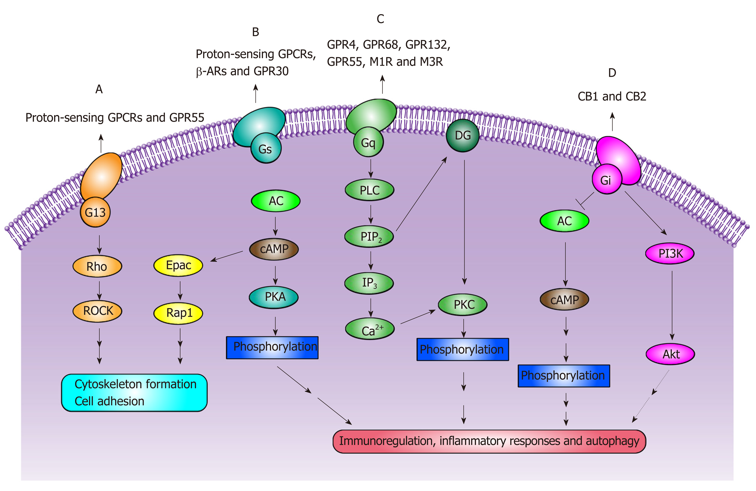
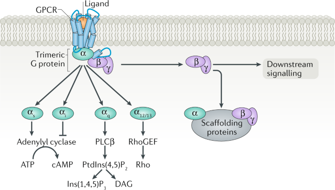
Diagram - G-protein coupled receptor common downstream activation pathways. In this diagram, lines ending in arrows (such as acting on adenylyl cyclase in Gs) indicate an increase in action, and lines ending in a 'T' shape (such as acting on adenyly cyclase in Gi) indicate a decrease in action. SimpleMed original by Thomas Burnell
As already stated earlier (slide 1.2.3), G protein-coupled receptors (GPCRs) form the largest class of drug targets in the human body.It is therefore appropriate to examine and understand them in some detail. The human genome contains genes for several hundred GPCRs.
GPCRdb contains reference data, interactive visualisation and experiment design tools for G protein-coupled receptors (GPCRs). GPCRdb curates sequence alignments, structures and receptor mutations from literature. Interactive diagrams visualise receptor residues (e.g. snakeplot and helix box plot) and relationships (e.g phylogenetic trees).
G Protein-Coupled Receptors Examples: Beta-adrenergic receptors that bind epinephrine; prostaglandin E2 receptors that bind inflammatory compounds called prostaglandins; and rhodopsin, which contains a photoreactive molecule called retinal that responds to light signals received by rod cells in the eye, are all examples of GPCRs.
These receptors are coupled to intracellular GTP-binding proteins (G-proteins). Once activated, G-proteins trigger the production of a variety of second messengers (e.g. cyclic AMP [cAMP], inositol triphosphate [IP3], diacylglycerol [DAG], etc.) helping to regulate a number of body functions ranging from sensation to growth to hormone release.
The metabotropic glutamate receptors (mGluRs) are family C G-protein-coupled receptors that participate in the modulation of synaptic transmission and neuronal excitability throughout the central nervous system. The mGluRs bind glutamate within a large ...
Background The G-protein coupled receptor (GPCR) superfamily consists of structurally similar proteins arranged into families (classes), and is one of the most abundant protein classes in the mammalian genome. 1-5 GPCRs undertake a plethora of essential physiological functions and are targets for numerous novel drugs. 4,5 Their ligands are structurally heterogenous, including natural ...










0 Response to "39 g protein coupled receptors diagram"
Post a Comment